Correlation between Bone Mineral Density Measured by Dual-Energy X-Ray Absorptiometry and Hounsfield Units Measured by Diagnostic CT in Lumbar Spine
- Affiliations
-
- 1Department of Neurosurgery, Seoul National University College of Medicine, Seoul, Korea.
- 2Neuroscience Research Institute, Seoul National University Medical Research Center, Seoul, Korea.
- 3Clinical Research Institute, Seoul National University Hospital, Seoul, Korea.
- 4Department of Medical Statistics, Seoul National University Boramae Medical Center, Seoul, Korea.
- 5Department of Neurosurgery, Seoul National University Boramae Medical Center, Seoul, Korea. ddolbae01@naver.com
- KMID: 2138353
- DOI: http://doi.org/10.3340/jkns.2013.54.5.384
Abstract
OBJECTIVE
Use of quantitative computed tomography (CT) to evaluate bone mineral density was suggested in the 1970s. Despite its reliability and accuracy, technical shortcomings restricted its usage, and dual-energy X-ray absorptiometry (DXA) became the gold standard evaluation method. Advances in CT technology have reduced its previous limitations, and CT evaluation of bone quality may now be applicable in clinical practice. The aim of this study was to determine if the Hounsfield unit (HU) values obtained from CT correlate with patient age and bone mineral density.
METHODS
A total of 128 female patients who underwent lumbar CT for back pain were enrolled in the study. Their mean age was 66.4 years. Among them, 70 patients also underwent DXA. The patients were stratified by decade of life, forming five age groups. Lumbar vertebrae L1-4 were analyzed. The HU value of each vertebra was determined by averaging three measurements of the vertebra's trabecular portion, as shown in consecutive axial CT images. The HU values were compared between age groups, and correlations of HU value with bone mineral density and T-scores were determined.
RESULTS
The HU values consistently decreased with increasing age with significant differences between age groups (p<0.001). There were significant positive correlations (p<0.001) of HU value with bone mineral density and T-score.
CONCLUSION
The trabecular area HU value consistently decreases with age. Based on the strong positive correlation between HU value and bone mineral density, CT-based HU values might be useful in detecting bone mineral diseases, such as osteoporosis.
MeSH Terms
Figure
-
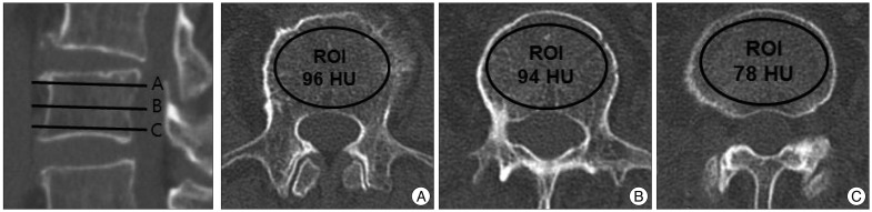
Fig. 1 Computed tomography (CT) scans of lumbar vertebra L3 illustrating the method of determining Hounsfield unit (HU) values by using a picture archiving and communication system (PACS). From a reconstructed sagittal image, we select three axial planes of interest : slice (A) is taken just inferior to the superior endplate, slice (B) is from the middle of the vertebral body, and slice (C) is taken just superior to the inferior endplate. The PACS program automatically calculates the mean HU value of the regions of interest which are marked with ellipses in the figure. The average of HU values from three axial cuts, which is 89 HU in this case, was used for the analysis.
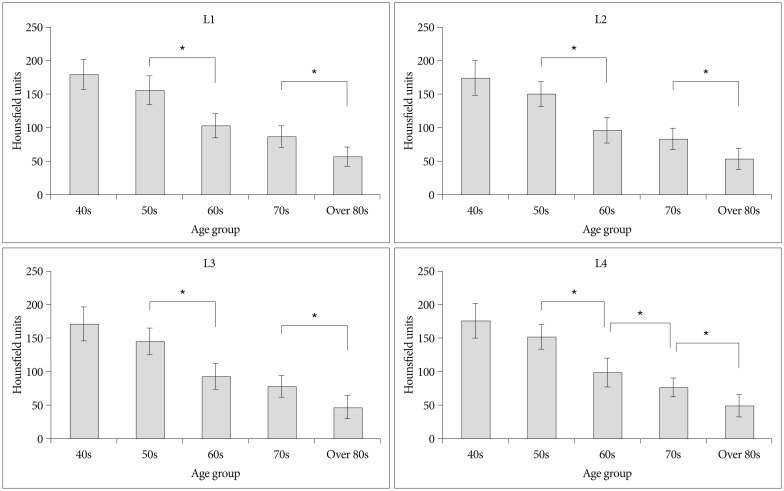
Fig. 2 Mean Hounsfield unit values among five decadal age groups show consistent decreases as age increases. *Groups which showed significant difference (p<0.05).

Fig. 3 Scatter plots showing correlations between Hounsfield unit values obtained from CT and T-scores obtained from dual-energy X-ray absorptiometry for lumbar vertebrae L1-L4. All had showed significant correlation coefficients (p<0.001).
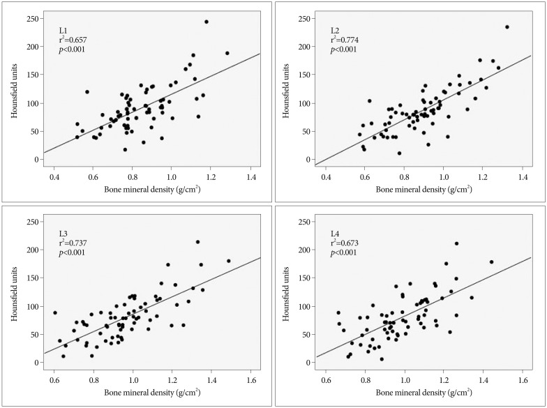
Fig. 4 Scatter plots showing correlations between Hounsfield unit values obtained from CT scans and bone marrow density obtained from dual-energy X-ray absorptiometry for lumbar vertebrae L1-4. All had significant correlation coefficients (p<0.001).
Cited by 1 articles
-
Machine Learning Model to Predict Osteoporotic Spine with Hounsfield Units on Lumbar Computed Tomography
Kyoung Hyup Nam, Il Seo, Dong Hwan Kim, Jae Il Lee, Byung Kwan Choi, In Ho Han
J Korean Neurosurg Soc. 2019;62(4):442-449. doi: 10.3340/jkns.2018.0178.
Reference
-
1. Adams JE. Quantitative computed tomography. Eur J Radiol. 2009; 71:415–424. PMID: 19682815.
Article2. Eswaran SK, Gupta A, Adams MF, Keaveny TM. Cortical and trabecular load sharing in the human vertebral body. J Bone Miner Res. 2006; 21:307–314. PMID: 16418787.
Article3. Goldman LW. Principles of CT and CT technology. J Nucl Med Technol. 2007; 35:115–128. quiz 129-130. PMID: 17823453.
Article4. Goo HW. CT radiation dose optimization and estimation : an update for radiologists. Korean J Radiol. 2012; 13:1–11. PMID: 22247630.
Article5. Keaveny TM, Hayes WC. A 20-year perspective on the mechanical properties of trabecular bone. J Biomech Eng. 1993; 115:534–542. PMID: 8302037.
Article6. Kim DH, Vaccaro AR. Osteoporotic compression fractures of the spine; current options and considerations for treatment. Spine J. 2006; 6:479–487. PMID: 16934715.
Article7. Kim KH, Lee K, Ko YJ, Kim SJ, Oh SI, Durrance DY, et al. Prevalence, awareness, and treatment of osteoporosis among Korean women : The Fourth Korea National Health and Nutrition Examination Survey. Bone. 2012; 50:1039–1047. PMID: 22366398.
Article8. Link TM, Koppers BB, Licht T, Bauer J, Lu Y, Rummeny EJ. In vitro and in vivo spiral CT to determine bone mineral density : initial experience in patients at risk for osteoporosis. Radiology. 2004; 231:805–811. PMID: 15105454.
Article9. Miyabara Y, Holmes D 3rd, Camp J, Miller VM, Kearns AE. Comparison of calibrated and uncalibrated bone mineral density by CT to DEXA in menopausal women. Climacteric. 2012; 15:374–381. PMID: 22175297.
Article10. Papadakis AE, Karantanas AH, Papadokostakis G, Damilakis J. Assessment of the morpho-densitometric parameters of the lumbar pedicles in osteoporotic and control women undergoing routine abdominal MDCT examinations. J Bone Miner Metab. 2011; 29:352–358. PMID: 20976512.
Article11. Papadakis AE, Karantanas AH, Papadokostakis G, Petinellis E, Damilakis J. Can abdominal multi-detector CT diagnose spinal osteoporosis? Eur Radiol. 2009; 19:172–176. PMID: 18641992.
Article12. Prevention and management of osteoporosis. World Health Organ Tech Rep Ser. 2003; 921:1–164. PMID: 15293701.13. Ross PD, Davis JW, Epstein RS, Wasnich RD. Pre-existing fractures and bone mass predict vertebral fracture incidence in women. Ann Intern Med. 1991; 114:919–923. PMID: 2024857.
Article
- Full Text Links
-
- Actions
-
Cited
- CITED
-
- Close
- Share
-
- Similar articles
-
- Assessment of Bone Mineral Density by Dual energy X-ray Absorptiometry in Korean Postemenopausal Women
- Cross-calibration of Bone Mineral Density between Two Different Dual X-ray Absorptiometry Systems: Hologic QDR 4500-A and Lunar EXPERT-XL
- Discordance between dual-energy X-ray absorptiometry bone mineral density and spinal computed tomography texture analysis: An investigation into low correlation rates
- Measurement of bone mineral density in osteoporotic fracture of the spine using dual energy X-ray absorptiometry
- Coincidence of Diagnosis of Osteoporosis at the Site of the Proximal Femur, Lumbar Spine and Distal Radius
