Laboratory Vestibular Function Testing
- Affiliations
-
- 1Department of Otolaryngology Head & Neck Surgery, Gachon University of College Medicine, Korea. han@gilhospital.com
- 2Department of Otolaryngology Head & Neck Surgery, Sungkyunkwan University School of Medicine, Korea. whehung@skku.edu
- KMID: 2137740
- DOI: http://doi.org/10.5124/jkma.2008.51.11.975
Abstract
- Basically laboratory vestibular function testing use the vestibular ocular reflex and vestibular spinal reflex like as bedside examination or outpatients' evaluation. Such vestibular laboratory testing can aid diagnosis and can be used to document an abnormality suspected at bedside evaluation. The ability to perform serial vestibular evaluations allows an assessment over time of patients who are undergoing treatment for dizziness or treatment with potentially ototoxic medication. Generally speaking, it includes spontaneous nystagmus, some kinds of evoked nystagmus, ocular eye movement testing, Caloric's testing, rotational chair testing, vestibular evoked myogenic potential, subjective visual vertical, posturography and so on. Those testing have been developed with biomedical engineering based on the proven scientific facts together.
Keyword
Figure
-

Figure 1 The results of positional test. It shows two types of horizontal canal benign positional paroxysmal vertigo. The ageotrophic horizontal direction changing positional nystagmus is shown on the top and it means the cupulolithiasis. Otherwise the geotrophic horizontal direction changing positional nystagmus is shown on the bottom and it means the canalithiasis. The lesion sides are right (top) and left (bottom) each.

Figure 2 The results of oculomotor testing. It shows saccadic (top) and pursuit (the middle) tests on left vestibulopathy patient, and then optokinetic (bottom) test on right vestibulopathy patients . Whole graphic data are affected by right beating (top and the middle) and left beating (bottom) spontaneous nystagmus.

Figure 3 The result of bithermal caloric test. It shows right acute vestibuopathy. The canal paresis and directional preponderance are 78%, 45% each. Visual fixation test is done on left side only and the value is not significant.
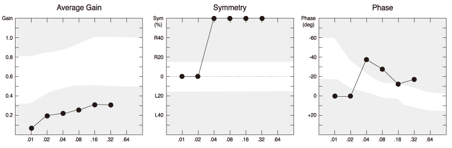
Figure 4 The results of sinusoidal harmonic acceleration test. It shows gain (top right), symmetry (top left), phase (bottom) on right acute vestibulopathy patients.

Figure 5 The results of step velocity test. It shows decreasing time constant on clockwise rotation (top) as 1 sec. Otherwise it is longer than that at counterclockwise rotation as 5 sec. It means the right acute vestibulopathy.

Figure 6 The results of dynamic posturograhy. The patient was fall down at condition 5 and 6 (right). Those condition means vestibular sensory organized condition (left).
Reference
-
1. Fetter Michael. Assessing vestibular function: which tests, when? J Neurol. 2000. 247:335–342.
Article2. Kingma Herman. Function tests of the otolith or statolith system. Curr Opin Neurol. 2006. 19:21–25.
Article3. Brandt Thomas, Strupp Michael. General vestibular testing. Clinical Neurophysiology. 2005. 116:406–426.
Article
- Full Text Links
-
- Actions
-
Cited
- CITED
-
- Close
- Share
-
- Similar articles
-
- Inferior Vestibular Neuritis: Absence of Vestibular Evoked Myogenic Potentials in the Presence of Normal Caloric Responses
- Vestibular Function Test in Old Age Patients with Vertebrobasilar Dolichoectasia
- The study of galvanic vestibular stimulation in patients of total unilateral vestibular loss
- The Principle and Methodology of Vestibular Evoked Myogenic Potential
- Value of Vestibular Function Tests for Diagnosis of Meniere's Disease
