J Korean Assoc Oral Maxillofac Surg.
2011 Apr;37(2):137-141. 10.5125/jkaoms.2011.37.2.137.
Injury of submandibular gland and lingual nerve as complication third molar tooth extraction in mandible : a case report
- Affiliations
-
- 1Department of Oral and Maxillofacial Surgery, Yeouido St. Mary's Hospital, The Catholic University of Korea, Seoul, Korea. justina@catholic.ac.kr
- KMID: 2137004
- DOI: http://doi.org/10.5125/jkaoms.2011.37.2.137
Abstract
- The extraction of an impacted third molar tooth is associated with many complications during the procedure and postoperative care. These complications include bleeding, swelling, pain, infection, as well as root fracture, proximal tooth injury, alveolar bone fracture, lingual nerve and inferior alveolar nerve injury etc. With the exception of a fractured root dislocation in the submandibular space, no direct submandibular gland injury related to extraction surgery has been reported until now. A 40 year old man visited the department of oromaxillofacial surgery at Yeouido St. Mary's Hospital for an extraction of the right mandible third molar. A partial third molar impaction was diagnosed by a clinical and radiographic examination. A surgical tooth extraction was practiced including buccal cortical bone osteotomy. During socket curettage, an encapsulated cyst-like lesion and a verified 3x3 cm neoplasm in the apically lingual direction were found during process of dissection. A biopsy confirmed that the neoplasm involved the submandibular gland and nerve trunk. This unusual anatomical organ injury during the surgical tooth extraction procedure is reported as a new complication during impacted third molar extraction.
MeSH Terms
Figure
-
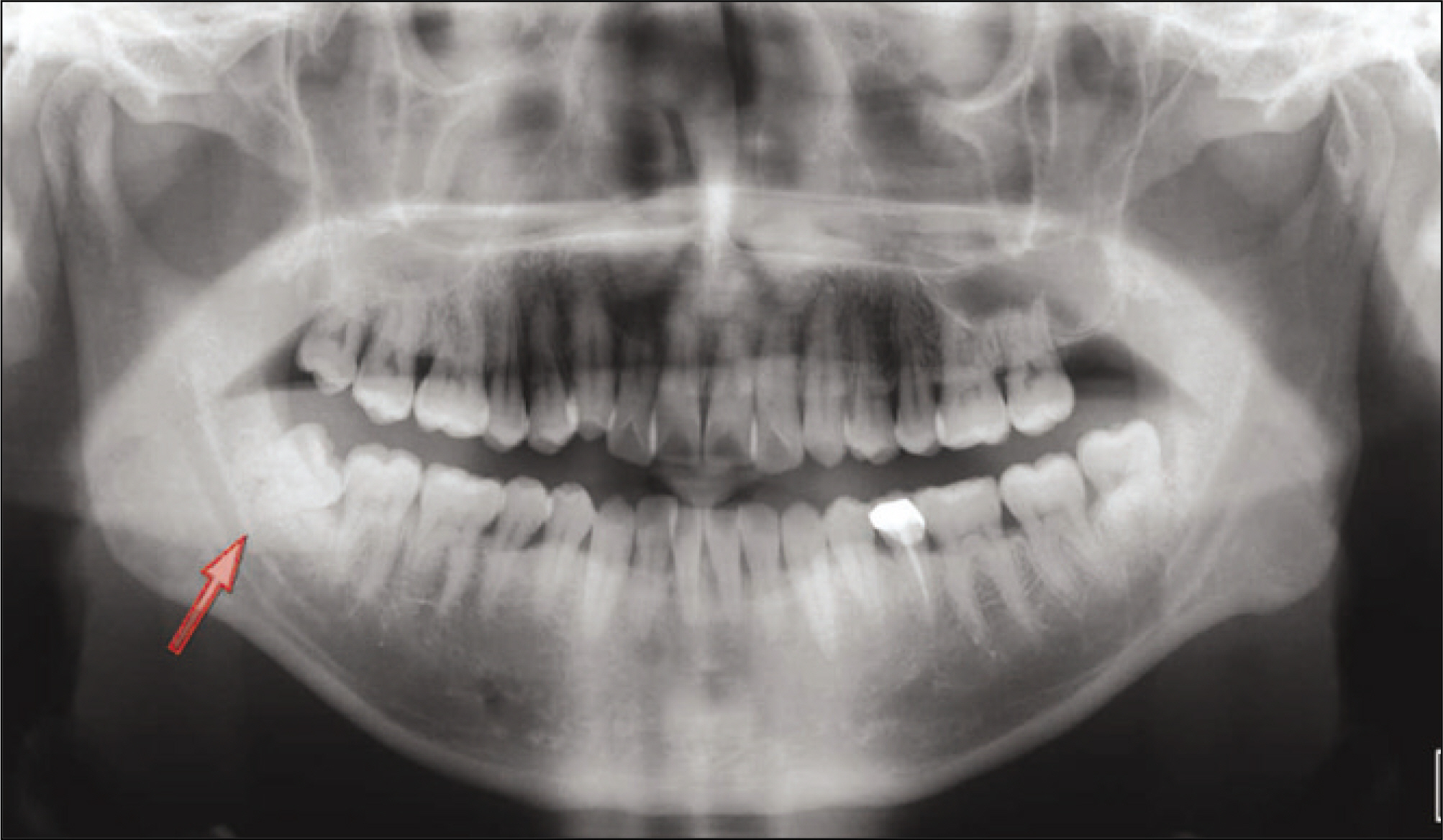
Fig. 1. Preoperative panoramaview. We diagnosed partial third molar impaction by radiographic examination.
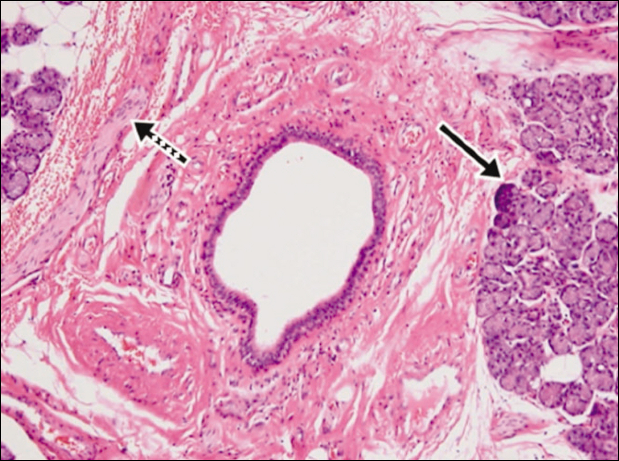
Fig. 2. Histologic finding.(H&E staining, original magnification x100) We confirmed that the neoplasm involved submandibular gland (line arrow) and nerve trunk (dot arrow).
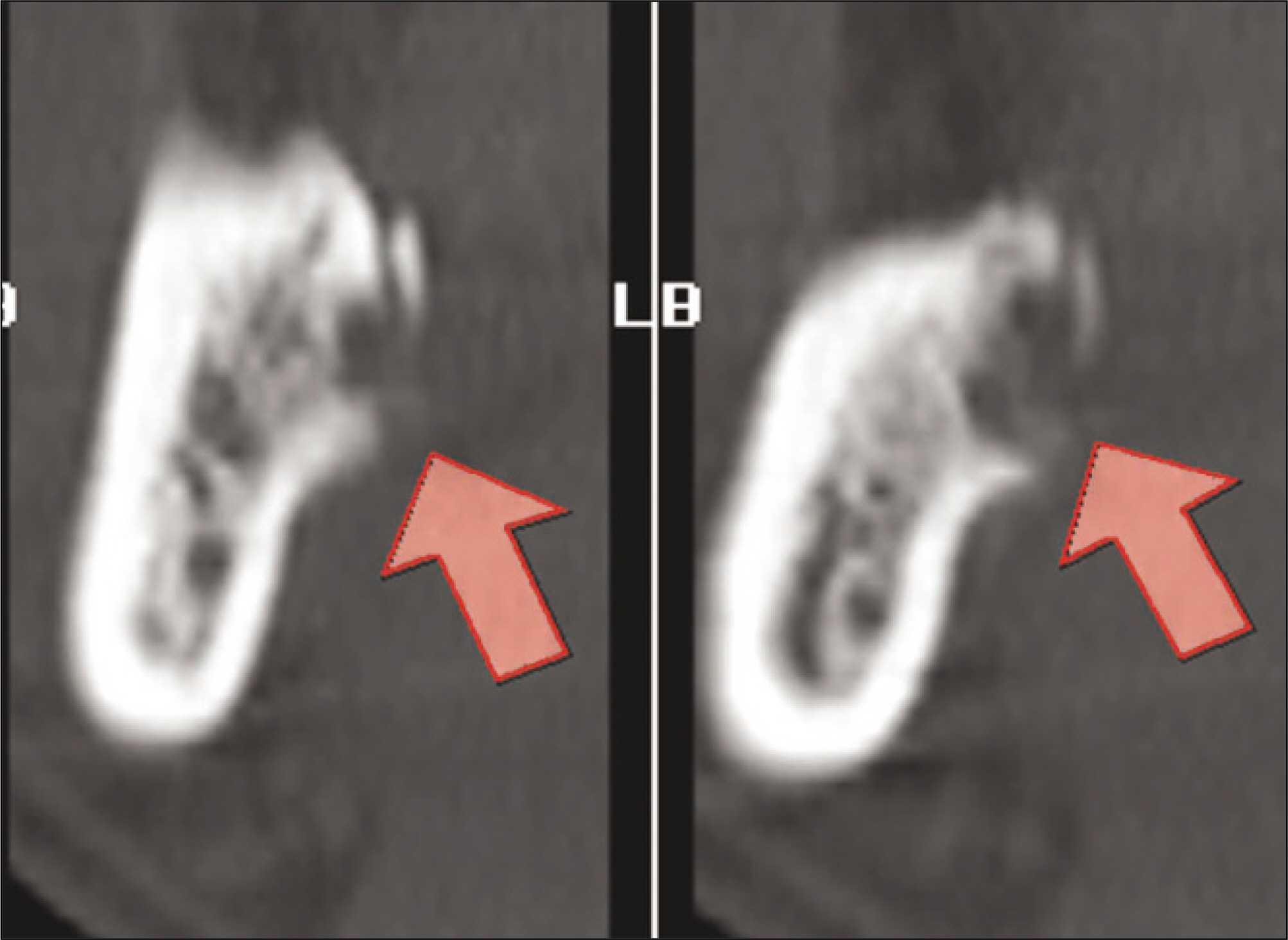
Fig. 3. Postoperative dental CT scan coronal view, non enhanced. We verified lingual bone breakage and partial resorption inner cortex of mandible.(CT: computed tomography)
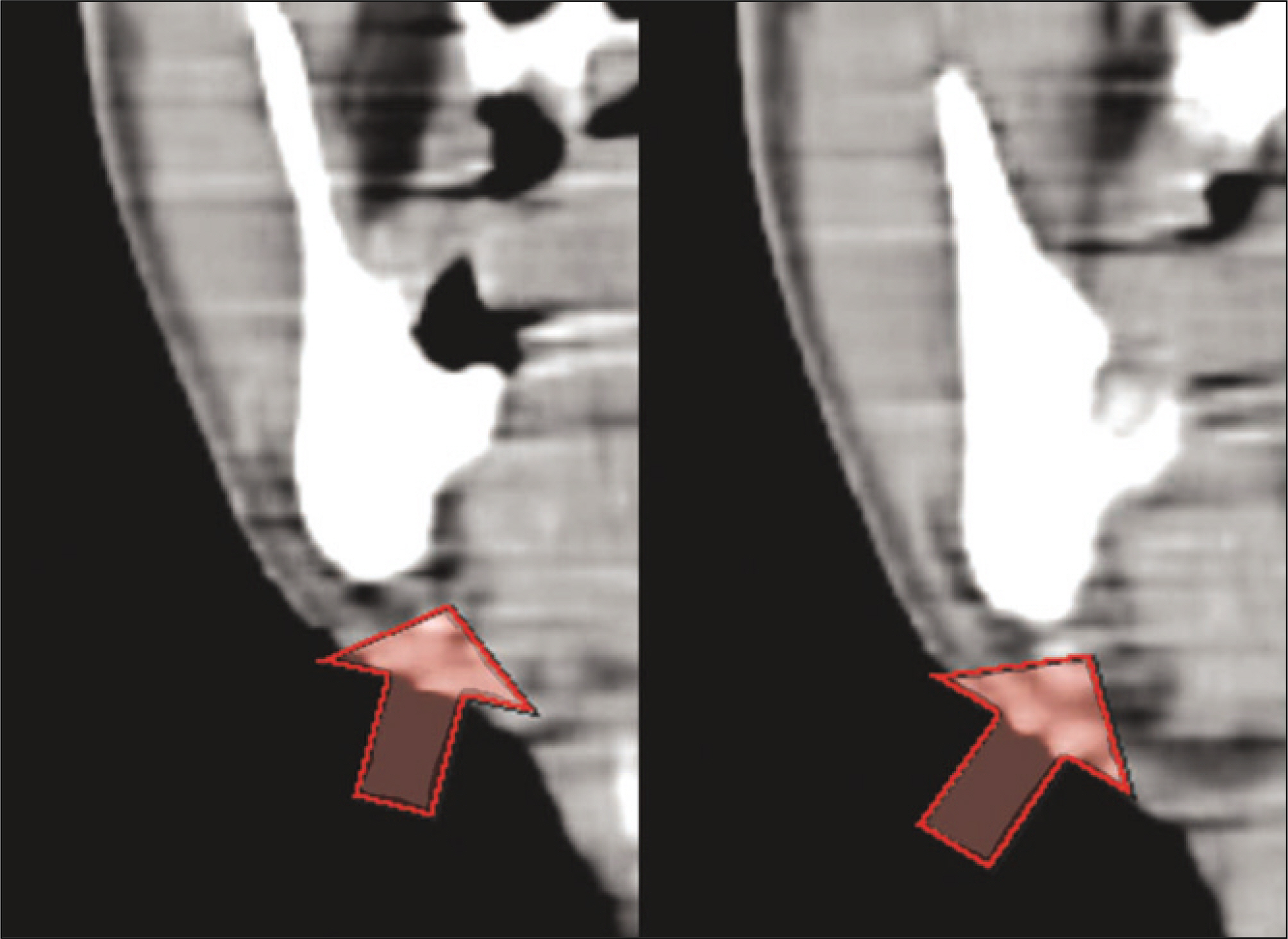
Fig. 4. Postoperative salivary gland CT scan coronal view, enhanced. We confirm submandibular gland close to lingual cortex bone and we diagnosed mild swelling and inflammatory change of submandibular gland due to tooth extraction injury.(CT: computed tomography)
Reference
-
References
1. Brauer HU. Unusual complications associated with third molar surgery: a systematic review. Quintessence Int. 2009; 40:565–72.2. Mason DA. Lingual nerve damage following lower third molar surgery. Int J Oral Maxillofac Surg. 1988; 17:290–4.
Article3. Song WS, Kim IK, Lee SH, Hwang YJ, Oh CY, Kim OJ. Complications of mandibular third molar extraction: two case reports of hyperventilation syndrome and displacement of roots into submandibular space. J Korean Assoc Maxillofac Plast Reconstr Surg. 2003; 25:568–72.4. Contar CM, de Oliveira P, Kanegusuku K, Berticelli RD, Azevedo-Alanis LR, Machado MA. Complications in third molar removal: a retrospective study of 588 patients. Med Oral Patol Oral Cir Bucal. 2010; 15:e74–8.
Article5. Blondeau F, Daniel NG. Extraction of impacted mandibular third molars: postoperative complications and their risk factors. J Can Dent Assoc. 2007; 73:325.6. Hiroyasu N, Kenichi S. Disturbance and regeneration of the inferior alveolar nerves. 1st ed.Seoul: Narae Publishing Co.;2001.7. Kiesselbach JE, Chamberlain JG. Clinical and anatomic observations on the relationship of the lingual nerve to the mandibular third molar region. J Oral Maxillofac Surg. 1984; 42:565–7.
Article8. Pogrel MA, Renaut A, Schmidt B, Ammar A. The relationship of the lingual nerve to the mandibular third molar region: an anatomic study. J Oral Maxillofac Surg. 1995; 53:1178–81.
Article9. Behnia H, Kheradvar A, Shahrokhi M. An anatomic study of the lingual nerve in the third molar region. J Oral Maxillofac Surg. 2000; 58:649–51. discussion 652–3.
Article10. Guerrissi JO, Taborda G. Endoscopic excision of the submandibular gland by an intraoral approach. J Craniofac Surg. 2001; 12:299–303.
Article11. Medeiros N, Gaffree G. Accidental displacement of inferior third molar into the lateral pharyngeal space: case report. J Oral Maxillofac Surg. 2008; 66:578–80.
Article12. Ertas U, Yaruz MS, Tozoglu S. Accidental third molar displacement into the lateral pharyngeal space. J Oral Maxillofac Surg. 2002; 60:1217.
Article13. Kiesselbach JE. The relationship of the lingual nerve to the mandibular third molar region: an anatomic study. J Oral Maxillofac Surg. 1995; 53:1181.
Article14. Philipsen HP, Takata T, Reichart PA, Sato S, Suei Y. Lingual and buccal mandibular bone depressions: a review based on 583 cases from a worldwide literature survey, including 69 new cases from Japan. Dentomaxillofac Radiol. 2002; 31:281–90.
Article15. Atieh MA. Diagnostic accuracy of panoramic radiography in determining relationship between inferior alveolar nerve and mandibular third molar. J Oral Maxillofac Surg. 2010; 68:74–82.
Article16. Sanfelice CM, da Costa FB, Reis So MV, Vier-Pelisser F, Souza Bier CA, Grecca FS. Effects of four instruments on coronal pre-enlargement by using cone beam computed tomography. J Endod. 2010; 36:858–61.
Article17. Isler SC, Demircan S, Soluk M, Cebi Z. Radiologic evaluation of an unusually sized complex odontoma involving the maxillary sinus by cone beam computed tomography. Quintessence Int. 2009; 40:533–5.18. Neugebauer J, Ritter L, Mischkowski RA, Dreiseidler T, Scherer P, Ketterle M, et al. Evaluation of maxillary sinus anatomy by conebeam CT prior to sinus floor elevation. Int J Oral Maxillofac Implants. 2010; 25:258–65.19. Chan HL, Leong DJ, Fu JH, Yeh CY, Tatarakis N, Wang HL. The significance of the lingual nerve during periodontal/implant surgery. J Periodontol. 2010; 81:372–7.
Article20. Tantanapornkul W, Okouchi K, Fujiwara Y, Yamashiro M, Maruoka Y, Ohbayashi N, et al. A comparative study of conebeam computed tomography and conventional panoramic radiography in assessing the topographic relationship between the mandibular canal and impacted third molars. Oral Surg Oral Med Oral Pathol Oral Radiol Endod. 2007; 103:253–9.
Article21. Ghaeminia H, Meijer GJ, Soehardi A, Borstlap WA, Mulder J, Berge SJ. Position of the impacted third molar in relation to the mandibular canal. Diagnostic accuracy of cone beam computed tomography compared with panoramic radiography. Int J Oral Maxillofac Surg. 2009; 38:964–71.
Article22. The Korean Association of Oral and Maxillofacial Surgeons. Textbook of oral and maxillofacial surgery. 2nd ed.Seoul: Dental and Medical Publishing Co.;2005.
- Full Text Links
-
- Actions
-
Cited
- CITED
-
- Close
- Share
-
- Similar articles
-
- Persistent lingual paresthesia caused by a displaced tooth fragment: a case report and literature review
- A CASE REPORT: STAFNE'S CYST IN THE ANTERIOR MANDIBLE
- Schwannoma Originating From the Lingual Nerve Radiologically Mimicking a Submandibular Gland Tumor
- Intraoral Excision of the Submandibular Gland
- A case report of a long-term abandoned torn lingual nerve injury repaired by collagen nerve graft induced by lower third molar extraction
