A Case of a Right Atrial and Inferior Vena Caval Thrombus Resembling a Right Atrial Myxoma
- Affiliations
-
- 1Division of Cardiology, Department of Internal Medicine, Ulsan Hospital, Ulsan, Korea.
- 2Division of Cardiology, Department of Internal Medicine, Pusan Paik Hospital, Inje University College of Medicine, Busan, Korea.
- KMID: 2135440
- DOI: http://doi.org/10.4250/jcu.2010.18.2.58
Abstract
- A right atrial and inferior vena caval thrombus in a structurally normal heart is a very rare condition. We report a case of such a thrombus in a 66-year-old woman. She was admitted to our hospital with recent onset dyspnea. Based on echocardiography, we suspected that she had myxoma. We performed an excision of a mass, which was found, by pathologic examination, to be an organized mural thrombus.
Keyword
MeSH Terms
Figure
-
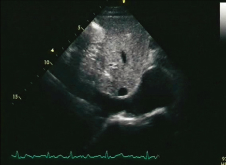
Fig. 1 Subcostal view of an echocardiogram shows a normal inferior vena cava and normal right atrium structures four years ago.

Fig. 2 Subcostal view of an echocardiogram shows two round masses having echolucent areas in the right atrium and dilated inferior vena cava (A). A transesophageal echocardiogram shows a mass with a stalk in the lower part of the right atrium (B).
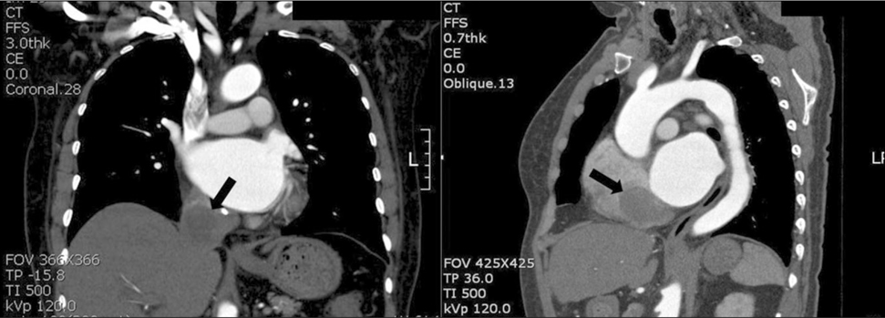
Fig. 3 A chest computed tomogram reveals two well-defined masses in the right atrium and inferior vena cava (arrows).
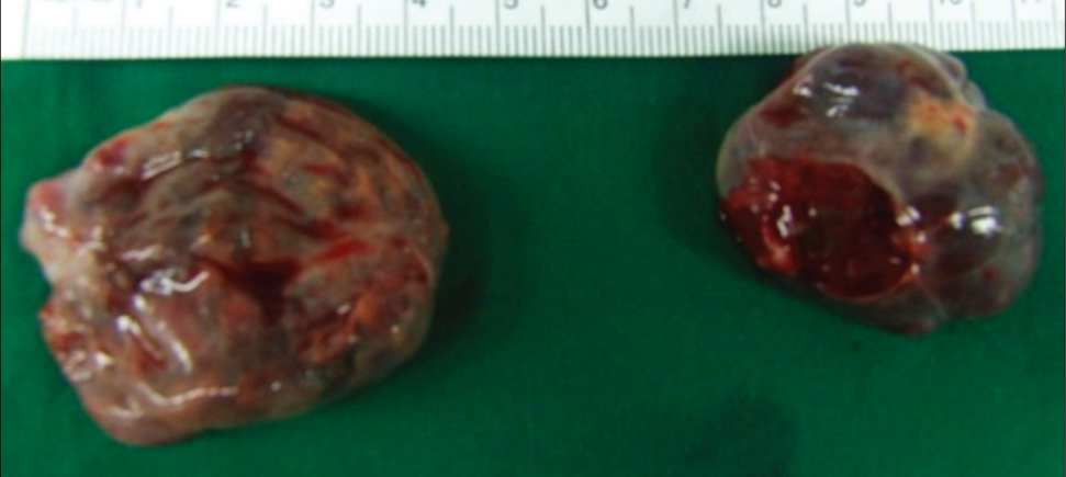
Fig. 4 Smooth-surfaced round masses were observed.
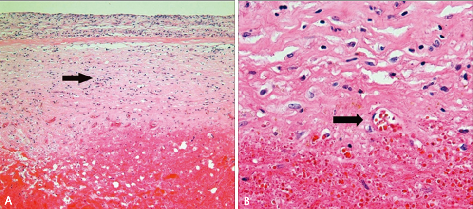
Fig. 5 The thrombus, which consists of platelets and fibrin with entrapped blood cells, is surrounded by fibrous tissue (arrow) (×100) (A). Arborizing vessels and fibroblasts are seen at the edge of the thrombus (arrow) (×400) (B).
Reference
-
1. Reynen K. Cardiac myxomas. N Engl J Med. 1995. 333:1610–1617.
Article2. Felner JM, Churchwell AL, Murpy DA. Right atrial thromboemboli: clinical, echocardiographic and pathophysiologic manifestations. J Am Coll Cardiol. 1984. 4:1041–1051.
Article3. Scheffel H, Baumueller S, Stolzmann P, Leschka S, Plass A, Alkadhi H, Schertler T. Atrial myxoma and thrombi: comparison of imaging features on CT. AJR Am J Roentgenol. 2009. 192:639–645.4. DeGroat TS, Parameswaran R, Popper PM, Kotler MN. Left ventricular thrombi in association with normal left ventricular wall motion in patients with malignancy. Am J Cardiol. 1985. 56:827–828.
Article5. Kim CH, Park DG, Kim SE, Yoon IS, Lee JH, Han KR, Oh DJ, Hong KS, Han SJ, Park WJ. A case of left ventricular thrombi in a patient with active ulcerative colitis and normal left ventricular systolic function. J Cardiovasc Ultrasound. 2006. 14:22–24.
Article6. Kim HJ, Yang JC, Kim SH, Chai JY, Jeon CH, Cha HS, Koh EM. A recurrent intracardiac thrombosis in a patient with Behet's disease. Korean J Med. 2005. 69:227–230.7. Panidis IP, Kotler MN, Mintz GS, Ross J. Clinical and echocardiographic features of right atrial masses. Am Heart J. 1984. 107:745–758.
Article8. Wartman WB, Hellerstein HK. The incidence of heart disease in 2,000 consecutive autopsies. Ann Intern Med. 1948. 28:41–65.
Article9. Pasierski TJ, Alton ME, Van Fossen DB, Pearson AC. Right atrial mobile thrombus: improved visualization by transesophageal echocardiography. Am Heart J. 1992. 123:802–803.
Article10. Porath A, Avnun L, Hirsch M, Ovsyshcher I. Right atrial thrombus and recurrent pulmonary amboli secondary to permanent cardiac pacing--a case report and short review of literature. Angiology. 1987. 38:627–630.
Article11. Mügge A, Gulba DC, Jost S, Daniel WG. Dissolution of a right atrial thrombus attached to pacemaker electrodes: usefulness of recombinant tissue-type plasminogen activator. Am Heart J. 1990. 119:1437–1439.
Article
- Full Text Links
-
- Actions
-
Cited
- CITED
-
- Close
- Share
-
- Similar articles
-
- Right Atrial Thrombus Mimicking Right Atrial Myxoma
- Hepatocellular carcinoma with atrial tumor thrombus presenting as myxoma: Resection under cardiopulmonary bypass
- Silent Left Large Atrial Myxoma: A Patient with Serial Electrocardiogram Variation
- Recurred Right Atrial Myxoma after Resection of Left Atrial Myxoma (Recurred Myxoma): A case report
- Case of Left Atrium Myxoma with Inferior Vena Caval Thrombus and Pulmonary Embolism Complicated with Budd-Chiari Syndrome
