Left Ventricular Rotation and Twist: Why Should We Learn?
- Affiliations
-
- 1Division of Functional Diagnostics, Department of Health Sciences, Osaka University Graduate School of Medicine, Osaka, Japan. nakatani@sahs.med.osaka-u.ac.jp
- KMID: 2135424
- DOI: http://doi.org/10.4250/jcu.2011.19.1.1
Abstract
- The left ventricle twists in systole storing potential energy and untwists (recoils) in diastole releasing the energy. Twist aids left ventricular ejection and untwist aids relaxation and ventricular filling. Therefore, rotation and torsion are important in cardiac mechanics. However, the methodology of their investigations is limited to invasive techniques or magnetic resonance imaging. With the advent of speckle tracking echocardiography, however, rotation and torsion (twist) become familiar to echocardiographers. In this review, I outline the mechanism and influencing factors of rotation and torsion with the anticipation of the routine use of these measurements in clinical practice.
Keyword
MeSH Terms
Figure
-
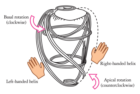
Fig. 1 Myocardial fiber orientation and direction of rotation. Myocardial fibers in the subepicardium helically run in a left-handed direction, fibers in the mid layer run circumferentially, and fibers in the subendocardium helically run in a right-handed direction.
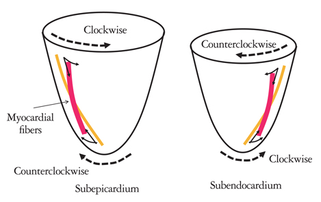
Fig. 2 Myocardial contraction and rotation. When myocardial fibers on the subepicardial side contract, clockwise rotational torque is produced at the base and counterclockwise rotational torque at the apex. When myocardial fibers on the subendocardial side contract, counterclockwise rotational torque is produced at the base and clockwise rotational torque at the apex.

Fig. 3 Opposite rotation at the base and apex. Subepicardial radius is larger than subendocardial radius (r2 > r1). Therefore, subepicardial rotational torque is larger than subendocardial rotational torque (R2 > R1).

Fig. 4 Tagged magnetic resonance imaging of a canine heart. Arrows at the apex mark the initial positions of two tags. By the end of isovolumic relaxation, tags have recoiled almost completely to their starting position indicating that recoil is largely an isovolumic event.11)
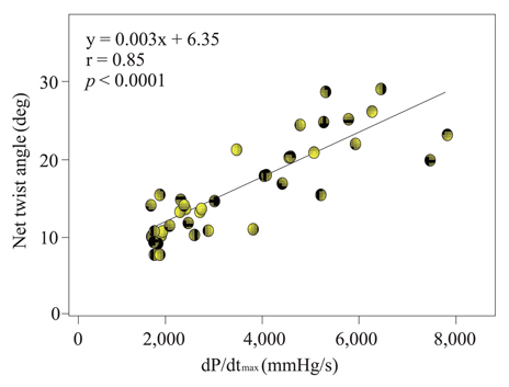
Fig. 5 Relationship between dP/dt and net twist angle.

Fig. 6 Hyper-rotation in the presence of subendocardial dysfunction. Apical rotation is shown as in Fig. 3. When there is subendocardial dysfunction, RT1' becomes smaller than RT1. Then, because of RT2 >> RT1', hyper-rotation is produced.
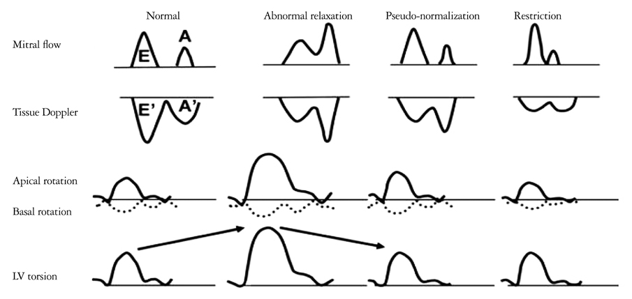
Fig. 7 Assessment of diastolic dysfunction based on mitral inflow, mitral annular velocity and left ventricular rotation and torsion. E: peak velocity of the early diastolic filling wave, A: peak velocity of the late diastolic filling wave, E': peak early diastolic annular velocity, A': peak late diastolic annular velocity.15)
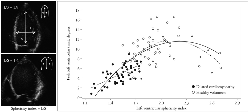
Fig. 8 Relationship between left ventricular shape and torsion. Left ventricular shape is expressed by the sphericity index that is the ratio of the long-axis diameter and the short-axis diameter in end-diastole (left). A parabolic relationship is found between the sphericity index and the torsion (twist angle) (right).20)
Cited by 1 articles
-
Estimating the Myocardium's Angle of Three-Dimensional Trajectory, Using the Tracking of Sequential Two-Dimensional Echocardiography Images
Mosayyeb Mobasheri, Manijhe Mokhtari-Dizaji, Faride Roshanali
J Cardiovasc Ultrasound. 2014;22(1):14-22. doi: 10.4250/jcu.2014.22.1.14.
Reference
-
1. Hansen DE, Daughters GT 2nd, Alderman EL, Ingels NB Jr, Miller DC. Torsional deformation of the left ventricular midwall in human hearts with intramyocardial markers: regional heterogeneity and sensitivity to the inotropic effects of abrupt rate changes. Circ Res. 1988. 62:941–952.
Article2. Gorman JH 3rd, Gupta KB, Streicher JT, Gorman RC, Jackson BM, Ratcliffe MB, Bogen DK, Edmunds LH Jr. Dynamic three-dimensional imaging of the mitral valve and left ventricle by rapid sonomicrometry array localization. J Thorac Cardiovasc Surg. 1996. 112:712–726.
Article3. Buchalter MB, Weiss JL, Rogers WJ, Zerhouni EA, Weisfeldt ML, Beyar R, Shapiro EP. Noninvasive quantification of left ventricular rotational deformation in normal humans using magnetic resonance imaging myocardial tagging. Circulation. 1990. 81:1236–1244.
Article4. Buchalter MB, Rademakers FE, Weiss JL, Rogers WJ, Weisfeldt ML, Shapiro EP. Rotational deformation of the canine left ventricle measured by magnetic resonance tagging: effects of catecholamines, ischaemia, and pacing. Cardiovasc Res. 1994. 28:629–635.
Article5. Mirro MJ, Rogers EW, Weyman AE, Feigenbaum H. Angular displacement of the papillary muscles during the cardiac cycle. Circulation. 1979. 60:327–333.
Article6. Notomi Y, Setser RM, Shiota T, Martin-Miklovic MG, Weaver JA, Popovic ZB, Yamada H, Greenberg NL, White RD, Thomas JD. Assessment of left ventricular torsional deformation by Doppler tissue imaging: validation study with tagged magnetic resonance imaging. Circulation. 2005. 111:1141–1147.
Article7. Notomi Y, Lysyansky P, Setser RM, Shiota T, Popovic ZB, Martin-Miklovic MG, Weaver JA, Oryszak SJ, Greenberg NL, White RD, Thomas JD. Measurement of ventricular torsion by two-dimensional ultrasound speckle tracking imaging. J Am Coll Cardiol. 2005. 45:2034–2041.
Article8. Sengupta PP, Tajik AJ, Chandrasekaran K, Khandheria BK. Twist mechanics of the left ventricle: principles and application. JACC Cardiovasc Imaging. 2008. 1:366–376.9. Henson RE, Song SK, Pastorek JS, Ackerman JJ, Lorenz CH. Left ventricular torsion is equal in mice and humans. Am J Physiol Heart Circ Physiol. 2000. 278:H1117–H1123.
Article10. Buckberg GD. Basic science review: the helix and the heart. J Thorac Cardiovasc Surg. 2002. 124:863–883.
Article11. Dong SJ, Hees PS, Siu CO, Weiss JL, Shapiro EP. MRI assessment of LV relaxation by untwisting rate: a new isovolumic phase measure of τ. Am J Physiol Heart Circ Physiol. 2001. 281:H2002–H2009.
Article12. Notomi Y, Popovic ZB, Yamada H, Wallick DW, Martin MG, Oryszak SJ, Shiota T, Greenberg NL, Thomas JD. Ventricular untwisting: a temporal link between left ventricular relaxation and suction. Am J Physiol Heart Circ Physiol. 2008. 294:H505–H513.
Article13. Dong SJ, Hees PS, Huang WM, Buffer SA Jr, Weiss JL, Shapiro EP. Independent effects of preload, afterload, and contractility on left ventricular torsion. Am J Physiol. 1999. 277:H1053–H1060.14. Owan TE, Hodge DO, Herges RM, Jacobsen SJ, Roger VL, Redfield MM. Trends in prevalence and outcome of heart failure with preserved ejection fraction. N Engl J Med. 2006. 355:251–259.
Article15. Park SJ, Miyazaki C, Bruce CJ, Ommen S, Miller FA, Oh JK. Left ventricular torsion by two-dimensional speckle tracking echocardiography in patients with diastolic dysfunction and normal ejection fraction. J Am Soc Echocardiogr. 2008. 21:1129–1137.
Article16. Wang J, Khoury DS, Yue Y, Torre-Amione G, Nagueh SF. Left ventricular untwisting rate by speckle tracking echocardiography. Circulation. 2007. 116:2580–2586.
Article17. Wang J, Khoury DS, Yue Y, Torre-Amione G, Nagueh SF. Preserved left ventricular twist and circumferential deformation, but depressed longitudinal and radial deformation in patients with diastolic heart failure. Eur Heart J. 2008. 29:1283–1289.
Article18. Notomi Y, Srinath G, Shiota T, Martin-Miklovic MG, Beachler L, Howell K, Oryszak SJ, Deserranno DG, Freed AD, Greenberg NL, Younoszai A, Thomas JD. Maturational and adaptive modulation of left ventricular torsional biomechanics: Doppler tissue imaging observation from infancy to adulthood. Circulation. 2006. 113:2534–2541.
Article19. Takeuchi M, Nakai H, Kokumai M, Nishikage T, Otani S, Lang RM. Age-related changes in left ventricular twist assessed by two-dimensional speckle-tracking imaging. J Am Soc Echocardiogr. 2006. 19:1077–1084.
Article20. Young AA, Kramer CM, Ferrari VA, Axel L, Reichek N. Three-dimensional left ventricular deformation in hypertrophic cardiomyopathy. Circulation. 1994. 90:854–867.
Article21. van Dalen BM, Kauer F, Vletter WB, Soliman OI, van der Zwaan HB, Ten Cate FJ, Geleijnse ML. Influence of cardiac shape on left ventricular twist. J Appl Physiol. 2010. 108:146–151.
Article22. Tanaka H, Oishi Y, Mizuguchi Y, Miyoshi H, Ishimoto T, Nagase N, Yamada H, Oki T. Contribution of the pericardium to left ventricular torsion and regional myocardial function in patients with total absence of the left pericardium. J Am Soc Echocardiogr. 2008. 21:268–274.
Article23. Chang SA, Kim HK, Kim YJ, Cho GY, Oh S, Sohn DW. Role of pericardium in the maintenance of left ventricular twist. Heart. 2010. 96:785–790.
Article
- Full Text Links
-
- Actions
-
Cited
- CITED
-
- Close
- Share
-
- Similar articles
-
- How Does the Left Ventricle Work? Ventricular Rotation as a New Index of Cardiac Performance
- Normal Left Ventricular Torsion Mechanics in Healthy Children: Age Related Changes of Torsion Parameters Are Closely Related to Changes in Heart Rate
- Left Ventricular Torsion Changes Post Kidney Transplantation
- Myocardial Rotation and Torsion in Child Growth
- Computerized Quantitative Analysis of Left Ventricular Wall Motion by 2-Dimensional Echocardiography
