J Cardiovasc Ultrasound.
2015 Mar;23(1):52-53. 10.4250/jcu.2015.23.1.52.
Basal Interventricular Septal Aneurysm in Rheumatic Mitral Stenosis
- Affiliations
-
- 1Department of Cardiology, King George's Medical University, Lucknow, Uttar Pradesh, India. dr.rajivkharwar@gmail.com
- KMID: 2135409
- DOI: http://doi.org/10.4250/jcu.2015.23.1.52
Abstract
- No abstract available.
Figure
-
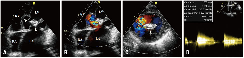
Fig. 1 Two dimensional transthoracic echocardiography. The presence of severe calcific mitral stenosis can be seen in apical four chamber as well as short axis view (arrow in A and C) with a mean and peak resting gradient of 30 mm Hg and 13 mm Hg respectively on continuous wave Doppler (D). The jet of the mitral stenosis is eccentrically directed towards the basal interventricular septum (color flow in B and C) likely responsible for the aneurysmal defect of the basal septum (arrowhead in A). Also note the marked dilatation of the LA. LA: left atria, LV: left ventricle, RA: right atria, RV: right ventricle.
Reference
-
1. Mohan JC, Kumar P. Cross-sectional echocardiographic diagnosis of a congenital aneurysm of the muscular interventricular septum. Int J Cardiol. 1992; 35:415–416.2. Kasprzak JD, Borkowski M, Rogowski W, Drozdz J, Krzemin´ska-Pakula M. A congenital complex including muscular interventricular septal aneurysm in an adult: case report and review of literature. Int J Cardiovasc Imaging. 2002; 18:25–30.3. Mohan JC, Nath LR. Aneurysmal deformity of the basal interventricular septum secondary to impinging turbulent transprosthetic eccentric flow jets. Indian Heart J. 2005; 57:258–260.
- Full Text Links
-
- Actions
-
Cited
- CITED
-
- Close
- Share
-
- Similar articles
-
- A Thrombus within an Interventricular Membranous Septal Aneurysm Leading to Cerebral Infarction: A Case Report
- A Giant Left Atrium in Rheumatic Mitral Stenosis
- Noninvasive Evaluation of Rheumatic Tricuspid Stenosis with Doppler and 2 Dimensional Echocardiography
- Rheumatic Carditis Associated with Mitral Stenosis
- Congenital Giant Aneurysm of Pulmonary Artery-Associated with Ventricular Septal Defect and Pulmonary Stenosis : A Case Report
