Double Chambered Right Ventricle with Ventricular Septal Defect in Adults: Case Series and Review of the Literature
- Affiliations
-
- 1Division of Cardiovascular Diseases, Mayo Clinic Arizona, Scottsdale, AZ, USA. sherifmoustafamd@yahoo.com
- 2Department of Cardiovascular Diseases, Prince Salman Heart Center, Riyadh, Saudi Arabia.
- 3Section of Pediatric Cardiology, University of Calgary, Calgary, AB, Canada.
- 4Division of Cardiovascular Diseases, University of Calgary, Calgary, AB, Canada.
- 5Department of Radiology, King Fahad Medical City, Riyadh, Saudi Arabia.
- KMID: 2135408
- DOI: http://doi.org/10.4250/jcu.2015.23.1.48
Abstract
- Double-chambered right ventricle (DCRV) is an uncommon congenital anomaly in which anomalous muscle bands divide the right ventricle into two chambers; a proximal high-pressure and distal low-pressure chamber. It may be associated with mid right ventricular obstruction. It is commonly associated with other congenital anomalies, most frequently perimembranous ventricular septal defect (PM-VSD). We herein present 5 adult patients with concomitant DCRV and PM-VSD who varied in their symptomatic presentations and the ways of management.
Keyword
Figure
-
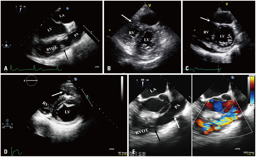
Fig. 1 A: Transesophageal echocardiogram mid esophageal view showing muscle band in the right ventricular outflow tract (arrow). B, C, and D: Transthoracic echocardiogram short axis views showing right ventricular muscle band (arrow). C: Posterior pericardial effusion was noted. E: Transesophageal echocardiogram mid esophageal view showing muscle band in the right ventricular outflow tract (left, black arrow) with color turbulence across (right). Pulmonic valve was shown (white arrow). LA: left atrium, LV: left ventricle, PA: pulmonary artery, RV: right ventricle, RVOT: right ventricular outflow tract.
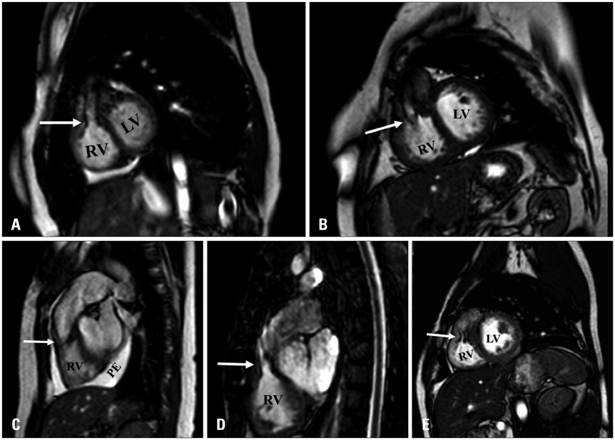
Fig. 2 A and B: Cardiac magnetic resonance: sagittal steady-state free precession views demonstrating prominent right ventricular outflow tract muscle bundles causing significant dephasing (arrow). C, D, and E: Cardiac magnetic resonance: sagittal steady-state free precession views demonstrating prominent right ventricular outflow tract muscle bundles without significant dephasing (arrow). C: Pericardial effusion was noted as well. LV: left ventricle, RV: right ventricle, PE: pericardial effusion.
Reference
-
1. Wong PC, Sanders SP, Jonas RA, Colan SD, Parness IA, Geva T, Van Praagh R, Spevak PJ. Pulmonary valve-moderator band distance and association with development of double-chambered right ventricle. Am J Cardiol. 1991; 68:1681–1686.2. Hindle WV Jr, Engle MA, Hagstrom JW. Anomalous right ventricular muscles: a clinicopathologic study. Am J Cardiol. 1968; 21:487–495.3. McElhinney DB, Chatterjee KM, Reddy VM. Double-chambered right ventricle presenting in adulthood. Ann Thorac Surg. 2000; 70:124–127.4. Nagashima M, Tomino T, Satoh H, Nakata T, Ohtani T, Saito H. Double-chambered right ventricle in adulthood. Asian Cardiovasc Thorac Ann. 2005; 13:127–130.5. Oliver JM, Garrido A, González A, Benito F, Mateos M, Aroca A, Sanz E. Rapid progression of midventricular obstruction in adults with double-chambered right ventricle. J Thorac Cardiovasc Surg. 2003; 126:711–717.6. Kottayil BP, Dharan BS, Pillai VV, Panicker VT, Gopalakrishnan SK, Jayakumar K. Surgical repair of double-chambered right ventricle in adulthood. Asian Cardiovasc Thorac Ann. 2011; 19:57–60.7. Chang RY, Kuo CH, Rim RS, Chou YS, Tsai CH. Transesophageal echocardiographic image of double-chambered right ventricle. J Am Soc Echocardiogr. 1996; 9:347–352.8. Kilner PJ, Sievers B, Meyer GP, Ho SY. Double-chambered right ventricle or sub-infundibular stenosis assessed by cardiovascular magnetic resonance. J Cardiovasc Magn Reson. 2002; 4:373–379.9. Darwazah AK, Eida M, Bader V, Khalil M. Surgical management of double-chambered right ventricle in adults. Tex Heart Inst J. 2011; 38:301–304.10. Pongiglione G, Freedom RM, Cook D, Rowe RD. Mechanism of acquired right ventricular outflow tract obstruction in patients with ventricular septal defect: an angiocardiographic study. Am J Cardiol. 1982; 50:776–780.
- Full Text Links
-
- Actions
-
Cited
- CITED
-
- Close
- Share
-
- Similar articles
-
- A Case of Double Chambered Right Ventricle with Congenital Right Ventricular True Diverticulum
- Two Cases of Double-Chambered Right Ventricle without Other Congenital Cardiac Anomalies
- Noonan Syndrome with Double-Chambered Right Ventricle and Atrial Septal Defect: 1 Case Report
- Spontaneously Healed Membranous Type Ventricular Septal Defect with Malaligned Interventricular Septal Wall and Double-Chambered Right Ventricle in a 56-Year-Old Patient
- Two-chambered right ventricle resulting from aberrant muscle bundles a case report
