Diffuse Unilateral Subacute Neuroretinitis in a Healthy Korean Male: The First Case Report in Korea
- Affiliations
-
- 1Institute of Vision Research, Department of Ophthalmology, Yonsei University College of Medicine, Seoul, Korea. sklee219@yuhs.ac
- 2Department of Ophthalmology, Catholic Kwandong University College of Medicine, International St.Mary's Hospital, Incheon, Korea.
- KMID: 2133316
- DOI: http://doi.org/10.3346/jkms.2015.30.3.346
Abstract
- A 52-yr-old male was referred for progressive visual loss in the left eye. The decimal best-corrected visual acuity (BCVA) was 0.01. Fundus examination revealed diffuse retinal pigment epithelial degeneration, focal yellow-white, infiltrative subretinal lesion with fuzzy border and a live nematode within the retina. Diffuse unilateral subacute neuroretinitis (DUSN) was diagnosed and the direct laser photocoagulation was performed to destroy the live nematode. During eight months after treatment, BCVA gradually improved to 0.2 along with the gradual restoration of outer retinal layers on SD-OCT. We report on the first case of DUSN in Korea. DUSN should be included in the differential diagnosis of unexplained unilateral visual loss in otherwise healthy subjects.
MeSH Terms
-
Animals
Blindness/diagnosis/parasitology
Eye Infections, Parasitic/diagnosis/parasitology/*therapy
Fundus Oculi
Humans
Laser Therapy/methods
Light Coagulation/methods
Male
Middle Aged
Nematoda/*pathogenicity
Republic of Korea
Retinal Pigment Epithelium/*parasitology/pathology
Retinitis/diagnosis/*parasitology/*therapy
Visual Acuity
Figure
-
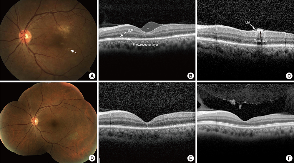
Fig. 1 Representative figures of the first Korean case with diffuse unilateral subacute neuroretinitis. A 52-yr-old male patient was referred to the tertiary ophthalmology clinic for gradual deterioration of vision in the left eye for the past 10 days. The best-corrected visual acuity (BCVA) in the left eye was 0.01 according to the decimal visual acuity chart. (A) Fundus of the left eye shows a yellow-white, infiltrative subretinal lesion with a whitish nematode (arrow) inferotemporal to the fovea. (B) Spectral domain optical coherence tomography (SD-OCT) of the left eye shows fluid accumulation in the epiretinal space of the fovea (asterisk), with diffuse disruption of external limiting membrane (ELM) and photoreceptor layer at fovea. (C) Cross-section SD-OCT image of a nematode beneath the internal limiting membrane (ILM). Under the diagnosis of diffuse unilateral subacute neuroretinitis, direct laser photocoagulation was done to eliminate the nematode. (D) After two weeks, infiltrative lesion was resolved. Note the pigmented area due to direct laser photocoagulation. (E) SD-OCT shows complete resolution of fluid at sub-ILM space and recovery of ELM. However, photoreceptor layer was not fully recovered. (F) At an 8-month follow-up visit, the BCVA of the left eye improved as 0.2. Diffuse disruption of photoreceptor disruption on SD-OCT was much recovered.
Reference
-
1. Gass JD, Braunstein RA. Further observations concerning the diffuse unilateral subacute neuroretinitis syndrome. Arch Ophthalmol. 1983; 101:1689–1697.2. Gass JD, Gilbert WR Jr, Guerry RK, Scelfo R. Diffuse unilateral subacute neuroretinitis. Ophthalmology. 1978; 85:521–545.3. Cortez RT, Ramirez G, Collet L, Giuliari GP. Ocular parasitic diseases: a review on toxocariasis and diffuse unilateral subacute neuroretinitis. J Pediatr Ophthalmol Strabismus. 2011; 48:204–212.4. de Souza EC, da Cunha SL, Gass JD. Diffuse unilateral subacute neuroretinitis in South America. Arch Ophthalmol. 1992; 110:1261–1263.5. de Souza EC, Nakashima Y. Diffuse unilateral subacute neuroretinitis. Report of transvitreal surgical removal of a subretinal nematode. Ophthalmology. 1995; 102:1183–1186.6. Goldberg MA, Kazacos KR, Boyce WM, Ai E, Katz B. Diffuse unilateral subacute neuroretinitis. Morphometric, serologic, and epidemiologic support for Baylisascaris as a causative agent. Ophthalmology. 1993; 100:1695–1701.7. Kazacos KR, Raymond LA, Kazacos EA, Vestre WA. The raccoon ascarid. A probable cause of human ocular larva migrans. Ophthalmology. 1985; 92:1735–1744.8. Cialdini AP, de Souza EC, Avila MP. The first South American case of diffuse unilateral subacute neuroretinitis caused by a large nematode. Arch Ophthalmol. 1999; 117:1431–1432.9. de Souza EC, Abujamra S, Nakashima Y, Gass JD. Diffuse bilateral subacute neuroretinitis: first patient with documented nematodes in both eyes. Arch Ophthalmol. 1999; 117:1349–1351.10. Garcia CA, Gomes AH, Garcia Filho CA, Vianna RN. Early-stage diffuse unilateral subacute neuroretinitis: improvement of vision after photocoagulation of the worm. Eye (Lond). 2004; 18:624–627.11. de Amorim Garcia Filho CA, Gomes AH, de A Garcia Soares AC, de Amorim Garcia CA. Clinical features of 121 patients with diffuse unilateral subacute neuroretinitis. Am J Ophthalmol. 2012; 153:743–749.12. Garcia CA, Gomes AH, Vianna RN, Souza Filho JP, Garcia Filho CA, Oréfice F. Late-stage diffuse unilateral subacute neuroretinitis: photocoagulation of the worm does not improve the visual acuity of affected patients. Int Ophthalmol. 2005; 26:39–42.13. Cortez R, Denny JP, Muci-Mendoza R, Ramirez G, Fuenmayor D, Jaffe GJ. Diffuse unilateral subacute neuroretinitis in Venezuela. Ophthalmology. 2005; 112:2110–2114.14. Naumann GO, Knorr HL. DUSN occurs in Europe. Ophthalmology. 1994; 101:971–972.15. Yuen VH, Chang TS, Hooper PL. Diffuse unilateral subacute neuroretinitis syndrome in Canada. Arch Ophthalmol. 1996; 114:1279–1282.16. Cai J, Wei R, Zhu L, Cao M, Yu S. Diffuse unilateral subacute neuroretinitis in China. Arch Ophthalmol. 2000; 118:721–722.17. Venkatesh P, Sarkar S, Garg S. Diffuse unilateral subacute neuroretinitis: report of a case from the Indian subcontinent and the importance of immediate photocoagulation. Int Ophthalmol. 2005; 26:251–254.18. Garcia Filho CA, Soares AC, Penha FM, Garcia CA. Spectral domain optical coherence tomography in diffuse unilateral subacute neuroretinitis. J Ophthalmol. 2011; 2011:285296.19. Tarantola RM, Elkins KA, Kay CN, Folk JC. Photoreceptor recovery following laser photocoagulation and albendazole in diffuse unilateral subacute neuroretinitis. Arch Ophthalmol. 2011; 129:669–671.20. Ament CS, Young LH. Ocular manifestations of helminthic infections: onchocersiasis, cysticercosis, toxocariasis, and diffuse unilateral subacute neuroretinitis. Int Ophthalmol Clin. 2006; 46:1–10.
- Full Text Links
-
- Actions
-
Cited
- CITED
-
- Close
- Share
-
- Similar articles
-
- Unilateral neuroretinitis and periparillary serous retinal detachment in cat-scratch disease
- A Case of Tubeculous Neuroretinitis
- Subacute Epiphyseal Osteomyelitis in a Child: A Case Report
- Recurrence of Subacute Thyroiditis: Report of Two Cases
- A Case of Subacute Thyroiditis Associated with Papillary Thyroid Carcinoma and Takayasu's Arteritis
