Multiple Giant Coronary Aneurysms Arising from Coronary Fistula to the Pulmonary Artery Revealed in Aorta CT Angiography
- Affiliations
-
- 1Department of Radiology, Dong-A University Hospital, Dong-A University College of Medicine, Busan, Korea.
- 2Department of Radiology, Kyungpook National University Hospital, Kyungpook National University College of Medicine, Daegu, Korea. jonglee@knu.ac.kr
- KMID: 2130946
- DOI: http://doi.org/10.3348/jksr.2015.73.6.398
Abstract
- Coronary fistula is a rare coronary abnormality through which blood drains into the cardiac chamber, great vessel or other vessels. In addition, giant aneurysm arising from coronary fistula is rare pathologic manifestation. Herein, we presented a rare case of multiple giant coronary artery aneurysms arising from coronary to pulmonary artery fistula in a 79-year-old woman presenting with sudden loss of consciousness. The aneurysms were detected using thoracic computed tomography angiography and consequently confirmed by invasive coronary angiography.
MeSH Terms
Figure
-
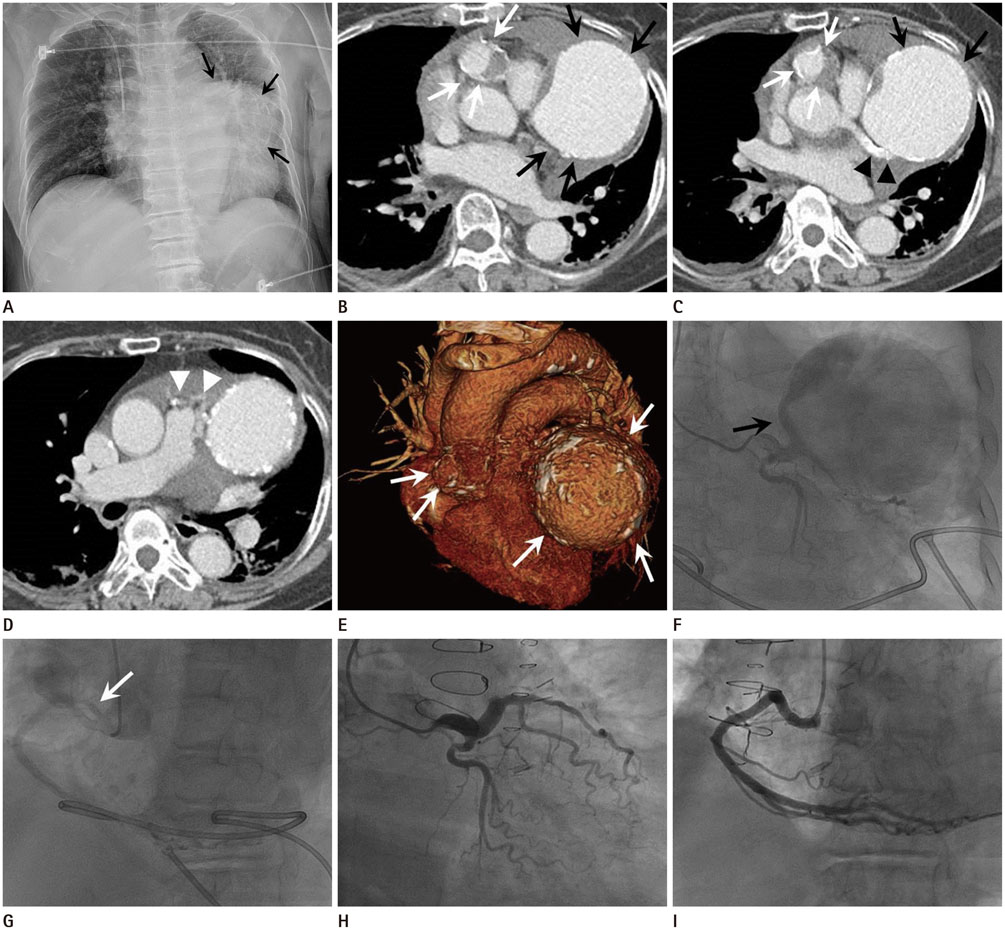
Fig. 1 A 79-year-old woman with multiple giant aneurysms arising from coronary to pulmonary artery fistula. A. Supine chest X-ray image shows an abnormal mass-like contour with calcified rim (black arrows) and obliterated left cardiac silhouette. B-E. Thoracic computed tomography angiography acquired during the venous phase. The axial scan shows an approximately 7-cm-sized contrast-filled lesion (black arrows) communicating with the left anterior descending coronary artery (black arrowheads on C). Another 2.5-cm-sized contrast-filled lesion (white arrows) with mural thrombus locates between the right coronary sinus and pulmonary trunk. Multiple tortuous anomalous vessels are noted anterior to the pulmonary trunk (white arrowheads on D), which suggest coronary fistulae. Three-dimensional volume-rendered image (E) shows schematic morphology of 2 giant coronary aneurysms (white arrows on E). F, G. Left coronary angiography demonstrates a giant coronary aneurysm originating from a branch (black arrow on F) of proximal left anterior descending coronary artery. Right coronary angiography shows another aneurysm from an anomalous branch (white arrow on G) of the right coronary sinus. H, I. Postoperative coronary angiography shows disappearance of previously noted coronary artery aneurysms and supplying vessels on both left and right coronary arteries.
Reference
-
1. Fernandes ED, Kadivar H, Hallman GL, Reul GJ, Ott DA, Cooley DA. Congenital malformations of the coronary arteries: the Texas Heart Institute experience. Ann Thorac Surg. 1992; 54:732–740.2. Pahlavan PS, Niroomand F. Coronary artery aneurysm: a review. Clin Cardiol. 2006; 29:439–443.3. Alcock R, Naoum C, Ng AC. Giant right coronary aneurysm: a case of mistaken identity. Eur Heart J. 2011; 32:2712.4. Wan S, LeClerc JL, Vachiery JL, Vincent JL. Cardiac tamponade due to spontaneous rupture of right coronary artery aneurysm. Ann Thorac Surg. 1996; 62:575–576.5. Mangukia CV. Coronary artery fistula. Ann Thorac Surg. 2012; 93:2084–2092.6. Edwards JE. Anomalous coronary arteries with special reference to arteriovenous-like communications. Circulation. 1958; 17:1001–1006.7. Said SA, el Gamal MI. Coronary angiographic morphology of congenital coronary arteriovenous fistulas in adults: report of four new cases and review of angiograms of fifteen reported cases. Cathet Cardiovasc Diagn. 1995; 35:29–35.8. Okita Y, Miki S, Kusuhara K, Ueda Y, Tahata T, Sakai T, et al. Aneurysm of coronary arteriovenous fistula presenting as a calcified mediastinal mass. Ann Thorac Surg. 1992; 54:771–773.9. Kim JY, Yoon YS, Pyun WB, Chang HJ, Choi SH, Lee DY, et al. A case of giant aneurysm of coronary arteriovenous fistula treated by percutaneous deployment of embolization coil. Korean Circ J. 1999; 29:1362–1365.10. Cao H, Ye L, Chan P, Fan H, Liu Z. Giant coronary artery aneurysm with fistula to the pulmonary artery complicated by frequent ventricular premature contractions: a case report. Medicine (Baltimore). 2015; 94:e530.
- Full Text Links
-
- Actions
-
Cited
- CITED
-
- Close
- Share
-
- Similar articles
-
- A case of coronary artery-pulmonary artery fistula communicated with aorto-pulmonary fistula via common channel detected by Multidetector row CT (MDCT) and coronary angiography
- Right Coronary Artery to Left Ventricular Fistula with a Giant Right Coronary Artery Aneurysm: A case report
- Dual Fistulas of Ascending Aorta and Coronary Artery to Pulmonary Artery
- Unusual Coronary Artery Fistula: Left Anterior Descending Coronary Artery - Left Ventricular Fistula Diagnosed by ECG-Gated Multi-Detector Row Coronary CT Angiography
- A Case of Coronary Arteriovenous Fistula Associated with Giant Coronary Artery Aneurysm
