Transplantation of a Horseshoe Kidney Found During Harvest Operation of a Cadaveric Donor: A Case Report
- Affiliations
-
- 1Department of Surgery, Soonchunhyang University College of Medicine, Seoul, Korea. backsa7@schmc.ac.kr
- 2Department of Urology, Soonchunhyang University College of Medicine, Seoul, Korea.
- 3Department of Internal Medicine, Soonchunhyang University College of Medicine, Seoul, Korea.
- KMID: 2129615
- DOI: http://doi.org/10.3346/jkms.2014.29.8.1166
Abstract
- A 34-yr-old female was diagnosed as being brain dead. Preoperative ultrasound revealed no abnormal focal lesions. However, the horseshoe kidney was identified during organ harvest. En bloc nephrectomy was performed. The kidney was divided at the midline of isthmus. The divided right kidney was discarded due to numerous arteries and veins. The divided left kidney was transplanted. After declamping, the kidney was well perfused and started clearing. Resistive index was 0.72. Glomerular filtration ratio was 84.69 mL/min on postoperative day 14. The horseshoe kidney can be successfully transplanted and could be a good solution for the shortage of organ donors.
Keyword
MeSH Terms
Figure
-

Fig. 1 Gross finding of the kidney. (A) En bloc nephrectomy was performed. The left kidney had single renal artery (dark arrow) and vein, whereas the right kidney had numerous arteries (blank arrows) and veins (blank pentagons). The isthmus contained a broad band of normal parenchyma. Both kidneys showed no variation in urinary collecting system (blank triangle is the right ureter, dark triangle is the left ureter). (B) In bench surgery, the kidney was divided at midline of isthmus, preserving its vasculature and urinary collecting system. The edges of the renal remnant (outer layer) were approximated using 2-0 absorbable polyglactin sutures after inner layer renorrhaphy. (C) The divided left kidney was transplanted at recipient's right iliac fossa. The renal artery was anastomosed to recipient's right external iliac artery, and the renal vein anastomosed to the right external iliac vein, end to side fashion. Divided isthmus involving small part of kidney low pole remained dark red color and achieved hemostasis after declamping.
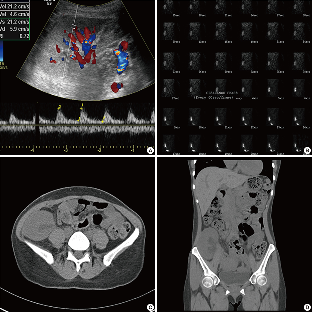
Fig. 2 Post-operative images and functions. (A) Renal doppler ultrasound at postoperative day 1. Size of the transplanted kidney was 11.2×4.3 cm. Parenchymal echogenicity was increased. RI index was 0.72. No abnormal pulsation wave was found. (B) DTPA-GFR results at postoperative 14 days. Normal concentration, but slight delayed excretion with calyectasis of transplanted kidney is seen. GFR of transplanted kidney was improved to 84.69 mL/min from 66.16 mL/min of postoperative 7 days. (C, D) Non-enhanced abdominal CT scan at postoperative one month. Transplanted kidney showed mild hydronephrosis with no definite evidence of radiopaque stone or dilatation of ureter.
Reference
-
1. Stroosma OB, Schurink GW, Smits JM, Kootstra G. Transplanting horseshoe kidneys: a worldwide survey. J Urol. 2001; 166:2039–2042.2. Zipitis CS, Augustine T, Tavakoli A, Surange R, Agrawal A, Riad HN. Horseshoe kidney transplantation. Surgeon. 2003; 1:160–163.3. Weizer AZ, Silverstein AD, Auge BK, Delvecchio FC, Raj G, Albala DM, Leder R, Preminger GM. Determining the incidence of horseshoe kidney from radiographic data at a single institution. J Urol. 2003; 170:1722–1726.4. Wein AJ, Kavoussi LR, Campbell MF, Walsh PC. Campbell-Walsh urology. 10th ed. Philadelphia: Elsevier Saunders;2012.5. Stroosma OB, Scheltinga MR, Stubenitsky BM, Kootstra G. Horseshoe kidney transplantation: an overview. Clin Transplant. 2000; 14:515–519.6. O'Brien J, Buckley O, Doody O, Ward E, Persaud T, Torreggiani W. Imaging of horseshoe kidneys and their complications. J Med Imaging Radiat Oncol. 2008; 52:216–226.7. Strauss S, Dushnitsky T, Peer A, Manor H, Libson E, Lebensart PD. Sonographic features of horseshoe kidney: review of 34 patients. J Ultrasound Med. 2000; 19:27–31.8. Stroosma OB, Smits JM, Schurink GW, de Boer J, Persijn GG, Kootstra G. Horseshoe kidney transplantation within the eurotransplant region: a case control study. Transplantation. 2001; 72:1930–1933.9. Goyal A, Gaitonde K, Sagade SN, Shah BV, Kamat MH. Transplantation of horseshoe kidney from living-related donors: report of two cases. Transplant Proc. 2003; 35:32–34.10. Salehipour M, Bahador A, Salahi H, Nikeghbalian S, Jalaeian H, Davari HR, Malek-Hosseini SA. Transplantation of a horseshoe kidney. Arch Iran Med. 2007; 10:239–241.11. Neville H, Ritchey ML, Shamberger RC, Haase G, Perlman S, Yoshioka T. The occurrence of Wilms tumor in horseshoe kidneys: a report from the National Wilms Tumor Study Group (NWTSG). J Pediatr Surg. 2002; 37:1134–1137.12. Lee SH, Bae MH, Choi SH, Lee JS, Cho YS, Joo KJ, Kwon CH, Park HJ. Wilms' tumor in a horseshoe kidney. Korean J Urol. 2012; 53:577–580.13. Kauffman HM, Cherikh WS, Cheng Y, Hanto DW, Kahan BD. Maintenance immunosuppression with target-of-rapamycin inhibitors is associated with a reduced incidence of de novo malignancies. Transplantation. 2005; 80:883–889.
