Multiple myeloma presenting with a maxillary lesion as the first sign
- Affiliations
-
- 1Department of Oral Medicine and Radiology, Navodaya Dental College and Hospital, Raichur, India. krksmile@gmail.com
- 2Department of Public Health Dentistry, Navodaya Dental College and Hospital, Raichur, India.
- KMID: 2116792
- DOI: http://doi.org/10.5624/isd.2015.45.1.55
Abstract
- Multiple myeloma is a clonal neoplastic proliferation of terminally differentiated B-lymphocytes involving the skeletal system in a multifocal fashion. Its oral manifestations are less common in the maxilla than in the mandible due to the lower amount of hemopoietic bone marrow in the maxilla. We report the case of a 50-year-old man who presented with a mass in the left maxillary alveolar region with tooth mobility. The mass had become enlarged after the teeth were extracted 15 days previously. Radiographs demonstrated multiple punched-out radiolucent lesions in the skull and pelvic region. Computed tomography images showed a soft tissue density mass in the left maxilla, eroding the floor and walls of the maxillary sinus. Although several analytical techniques were used to characterize the lesion, it was finally confirmed as multiple myeloma through immunohistochemistry.
Keyword
MeSH Terms
Figure
-
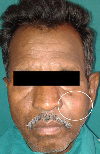
Fig. 1 An extraoral photograph shows diffuse swelling in the left middle third of the face.
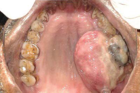
Fig. 2 An intraoral photograph shows a sessile mass in the left maxillary alveolus.
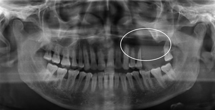
Fig. 3 A panoramic radiograph shows an osteolytic lesion in the left posterior maxilla, with resorption of the hard palate and the floor of the maxillary sinus.
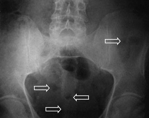
Fig. 4 An antero-posterior pelvic radiograph shows multiple punchedout radiolucent lesions.
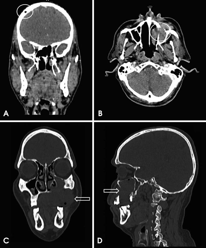
Fig. 5 A. A coronal computed tomography image of the skull shows punched-out radiolucent lesions. B. An axial computed tomography image shows a soft tissue density mass in the left maxilla, eroding the medial and lateral walls. C. A coronal computed tomography image shows erosion of the left maxillary sinus floor and the maxillary alveolus. D. A sagittal computed tomography image shows erosion of the anterior wall of the maxillary sinus with involvement of the premaxillary tissues.
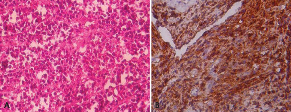
Fig. 6 Photomicrographs show the proliferating sheets of atypical plasma cells (A, H&E stain, 200×) and the immunohistochemically positive cells for CD138 (B, 200×).
Reference
-
1. Sharma V, Sharma A. Punched-out lesions in skull. Multiple myeloma. N Z Med J. 2010; 123:81–82.2. Lee SH, Huang JJ, Pan WL, Chan CP. Gingival mass as the primary manifestation of multiple myeloma: report of two cases. Oral Surg Oral Med Oral Pathol Oral Radiol Endod. 1996; 82:75–79.3. Bruce KW, Royer RQ. Multiple myeloma occurring in the jaws: a study of 17 cases. Oral Surg Oral Med Oral Pathol. 1953; 6:729–744.4. Witt C, Borges AC, Klein K, Neumann HJ. Radiographic manifestations of multiple myeloma in the mandible: a retrospective study of 77 patients. J Oral Maxillofac Surg. 1997; 55:450–455.
Article5. Raubenheimer EJ, Lello GE, Dauth J, Fayman MS, Dvornak N, Senekal JC. Multiple myeloma presenting as localized expansile jaw tumour. Int J Oral Maxillofac Surg. 1988; 17:382–385.
Article6. Raley LL, Granite EL. Plasmacytoma of the maxilla: report of case. J Oral Surg. 1977; 35:497–500.7. Currie WJ, Hill RR, Keshani DK. An unusual case of maxillary tuberosity enlargement. Br Dent J. 1994; 177:60–62.8. Albright RL, Finkelman A, Doner JM, Beaubien R. Multiple myeloma with manifestation of a maxillary bony lesion and plasmacytoma. Oral Surg Oral Med Oral Pathol. 1968; 26:167–172.
Article9. Lesmes D, Laster Z. Plasmacytoma in the temporomandibular joint: a case report. Br J Oral Maxillofac Surg. 2008; 46:322–324.
Article10. Bird JM, Owen RG, D'Sa S, Snowden JA, Pratt G, Ashcroft J, et al. Guidelines for the diagnosis and management of multiple myeloma 2011. Br J Haematol. 2011; 154:32–75.
Article11. Kyle RA, Gertz MA, Witzig TE, Lust JA, Lacy MQ, Dispenzieri A, et al. Review of 1027 patients with newly diagnosed multiple myeloma. Mayo Clin Proc. 2003; 78:21–33.
Article12. Lambertenghi-Deliliers G, Bruno E, Cortelezzi A, Fumagalli L, Morosini A. Incidence of jaw lesions in 193 patients with multiple myeloma. Oral Surg Oral Med Oral Pathol. 1988; 65:533–537.13. Nau KC, Lewis WD. Multiple myeloma: diagnosis and treatment. Am Fam Physician. 2008; 78:853–859.14. Ashcroft AJ, Davies FE, Morgan GJ. Aetiology of bone disease and the role of bisphosphonates in multiple myeloma. Lancet Oncol. 2003; 4:284–292.
Article15. Mozaffari E, Mupparapu M, Otis L. Undiagnosed multiple myeloma causing extensive dental bleeding: report of a case and review. Oral Surg Oral Med Oral Pathol Oral Radiol Endod. 2002; 94:448–453.
Article16. Witt C, Borges AC, Klein K, Neumann HJ. Radiographic manifestations of multiple myeloma in the mandible: a retrospective study of 77 patients. J Oral Maxillofac Surg. 1997; 55:450–453.
Article17. Miller CD, Goltry RR, Shenasky JH. Multiple myeloma involving the mandible. Report of a case. Oral Surg Oral Med Oral Pathol. 1969; 28:603–609.18. Pisano JJ, Coupland R, Chen S, Miller AS. Plasmacytoma of the oral cavity and jaws: a clinicopathologic study of 13 cases. Oral Surg Oral Med Oral Pathol Oral Radiol Endod. 1997; 83:265–271.19. Lae ME, Vencio EF, Inwards CY, Unni KK, Nascimento AG. Myeloma of the jaw bones: a clinicopathologic study of 33 cases. Head Neck. 2003; 25:373–381.
Article20. Durie BG, Waxman AD, D'Agnolo A, Williams CM. Whole body (18)F-FDG PET identifies high-risk myeloma. J Nucl Med. 2002; 43:1457–1463.21. Schirrmeister H, Buck AK, Bergmann L, Reske SN, Bommer M. Positron emission tomography (PET) for staging of solitary plasmacytoma. Cancer Biother Radiopharm. 2003; 18:841–845.
Article22. Wood NK, Goaz PW. Differential diagnosis of oral and maxillofacial lesions. 5th ed. St. Louis: Mosby;1997. p. 387–389.23. Zhao XJ, Sun J, Wang YD, Wang L. Maxillary pain is the first indication of the presence of multiple myeloma: a case report. Mol Clin Oncol. 2014; 2:59–64.
Article
