Unusually large erupted complex odontoma: A rare case report
- Affiliations
-
- 1Department of Oral Medicine and Radiology, ITS Centre for Dental Studies and Research, Murad Nagar, Ghaziabad, Uttar Pradesh, India. drshivanandb@yahoo.com
- KMID: 2116791
- DOI: http://doi.org/10.5624/isd.2015.45.1.49
Abstract
- Odontomas are nonaggressive, hamartomatous developmental malformations composed of mature tooth substances and may be compound or complex depending on the extent of morphodifferentiation or on their resemblance to normal teeth. Among them, complex odontomas are relatively rare tumors. They are usually asymptomatic in nature. Occasionally, these tumors become large, causing bone expansion followed by facial asymmetry. Odontoma eruptions are uncommon, and thus far, very few cases of erupted complex odontomas have been reported in the literature. Here, we report the case of an unusually large, painless, complex odontoma located in the right posterior mandible.
Keyword
Figure
-

Fig. 1 Extraoral photograph shows gross facial asymmetry.

Fig. 2 Intraoral photograph shows a swelling with an ulcer on the buccal mucosa.
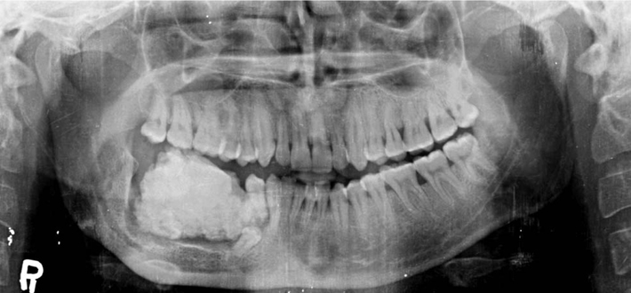
Fig. 3 Panoramic radiograph shows the lesion as a well-defined radiopacity surrounded by a radiolucent halo with secondary inferior displacement and oblique horizontal impaction of the right mandibular second premolar.
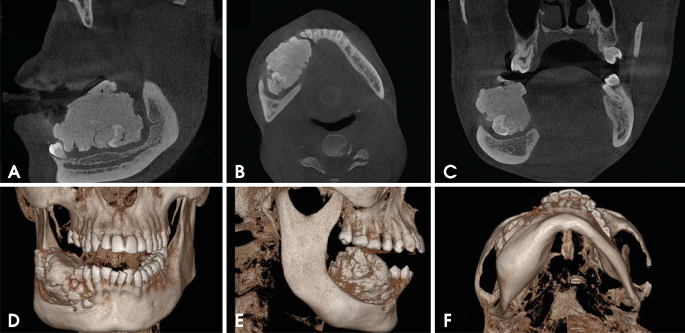
Fig. 4 A. Sagittal section of cone-beam computed tomography (CBCT) shows a dense homogeneous ground-glass radiopaque matrix with a horizontally aligned distinct molar-like tooth, adherent to the posteroinferior third of the mass. B. Axial section of CBCT shows the similar features of the sagittal section with a buccolingual expansion of the mandible. C. Coronal section of CBCT shows the similar features of the axial and sagittal sections with a buccolingual expansion and a tooth in the inferior portion of the lesion. Three-dimensional images of the lesion in the anteroposterior view (D) lateral view (E) submental view (F) show the expansion of cortical bones.

Fig. 5 Excised lesion weighing 43.5 g

Fig. 6 A. Ground section of complex odontoma (40×). B. Hematoxylin and eosin stained at 10× magnification. C. Hematoxylin and eosin stained at 40× magnification
Cited by 1 articles
-
Giant complex odontoma in the posterior mandible: A case report and literature review
Jong Chan Park, Ji Ho Yang, Sung Youn Jo, Bong Chul Kim, Jun Lee, Wan Lee
Imaging Sci Dent. 2018;48(4):289-293. doi: 10.5624/isd.2018.48.4.289.
Reference
-
1. Reichart P, Philipsen HP. Odontogenic tumours and allied lesions. London: Quintessence Pub;2004. p. 141–147.2. Vengal M, Arora H, Ghosh S, Pai KM. Large erupting complex odontoma: a case report. J Can Dent Assoc. 2007; 73:169–173.3. Dua N, Kapila R, Trivedi A, Mahajan S, Gupta SD. An unusual case of erupted composite complex odontoma. J Dent Sci Res. 2011; 2:1–5.4. Serra-Serra G, Berini-Aytes L, Gay-Escoda C. Erupted odontomas: a report of three cases and review of literature. Med Oral Patol Oral Cir Bucal. 2009; 14:E299–E303.5. Garcia-Consuegra L, Junquera LM, Albertos JM, Rodriguez . Odontomas. A clinical-histological and retrospective epidemiological study of 46 cases. Med Oral. 2000; 5:367–372.6. Mupparapu M, Singer SR, Rinaggio J. Complex odontoma of unusual size involving the maxillary sinus: report of a case and review of CT and histopathologic features. Quintessence Int. 2004; 35:641–645.7. De Visscher JG, Güven O, Elias AG. Complex odontoma in the maxillary sinus. Report of 2 cases. Int J Oral Surg. 1982; 11:276–280.8. Hitchin AD. The aetiology of the calcified composite odontomes. Br Dent J. 1971; 130:475–482.
Article9. Budnick SD. Compound and complex odontomas. Oral Surg Oral Med Oral Pathol. 1976; 42:501–506.
Article10. Kaur GA, Sivapathasundharam B, Berkovitz BK, Radhakrishnan RA. An erupted odontoma associated with pigmentation: a histogenetic and histological perspective. Indian J Dent Res. 2012; 23:699.
Article11. Philipsen HP, Reichart PA. Classification of odontogenic tumours. A historical review. J Oral Pathol Med. 2006; 35:525–529.
Article12. White SC, Pharoah MJ. Oral radiology: principles and interpretation. 6th ed. St. Louis: Mosby Elsevier;2010. p. 380–383.13. Wood NK, Goaz PW. Differential diagnosis of oral maxillofacial lesions. 5th ed. St. Louis: Mosby;2007. p. 492.14. Kodali RM, Venkat Suresh B, Ramanjaneya Raju P, Vora SK. An unusual complex odontoma. J Maxillofac Oral Surg. 2010; 9:314–317.
Article15. Ragalli CC, Ferreria JL, Blasco F. Large erupting complex odontoma. Int J Oral Maxillofac Surg. 2000; 29:373–374.
Article
