Plain Radiograph Analysis of the Distal Humerus Posterior Bowing That May Affect Interlocking Intramedullary Nailing for Humerus Shaft Fracture
- Affiliations
-
- 1Department of Orthopaedic Surgery, Sanggye Paik Hospital, Inje University of College of Medicine, Seoul, Korea. bkh26@hanmail.net
- KMID: 2106735
- DOI: http://doi.org/10.4055/jkoa.2015.50.1.31
Abstract
- PURPOSE
No research on posterior bowing of the distal humerus in the sagittal plane requiring evaluation during performance of intramedullary nailing has been reported in Korea. This study is designed to evaluate the location and angle of distal humeral posterior bowing in the sagittal plane through analysis of true lateral radiographs of humerus and discusses key points when performing intramedullary nailing.
MATERIALS AND METHODS
A retrospective study was conducted on 99 people with a simple lateral radiograph of the humerus and the authors analyzed total length of humerus, the angle and location of maximum posterior bowing in the distal shaft of the humerus.
RESULTS
The mean length of the humerus was 319.7 mm, and the mean angle of the distal posterior bowing was 8.8 degrees. The mean point of posterior bowing was 221.6 mm from the proximal end, which was 69.3% of the total length of the humerus.
CONCLUSION
The average posterior angulation of humerus existed at the point of 69.3% from the proximal humerus. Careful assessment is needed during intramedullary nailing in order to prevent complications.
Figure
-
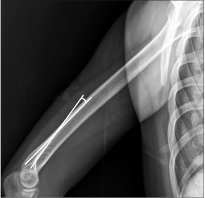
Figure 1 Measurement of posterior angulation of the distal humerus.

Figure 2 Measurement of the distance between the tip of the proximal humerus and the maximal point of posterior angulation of the distal humerus.

Figure 3 (A) True lateral radiograph of elbow before surgery shows 10 degrees of posterior angulation and 16 mm width of intramedullary canal. The fracture occurred approximately 70 mm proximally from the point of maximal posterior angulation. (B) Postoperative radiograph shows anterior angulation and displacement of the fracture site (arrow).

Figure 4 (A) Insertion of an intramedullary nail should be done with caution when site of fracture (imaginary dashed line) is at the distal humeral shaft. Lateral view of the humeral shaft shows that the intramedullary nail impinges with the posterior cortex of the distal humeral shaft. (B) Immediate postoperative radiograph shows posterior cortical breakage (arrow) resulting from impingement of the intramedullary nail with the distal humeral shaft due to posterior angulation of the humeral shaft.
Reference
-
1. Crates J, Whittle AP. Antegrade interlocking nailing of acute humeral shaft fractures. Clin Orthop Relat Res. 1998; 350:40–50.
Article2. Lin J. Treatment of humeral shaft fractures with humeral locked nail and comparison with plate fixation. J Trauma. 1998; 44:859–864.
Article3. Akpinar F, Aydinlioğlu A, Tosun N, Doğan A, Tuncay I, Unal O. A morphometric study on the humerus for intramedullary fixation. Tohoku J Exp Med. 2003; 199:35–42.
Article4. Park SR, Lee TJ, Kim RS, Moon KH, You DS. Result of interlocking intramedullary nailing for humeral shaft fracture evaluation of post-operative shoulder function. J Korean Fract Soc. 2007; 20:166–171.
Article5. Verdano MA, Pellegrini A, Schiavi P, Somenzi L, Concari G, Ceccarelli F. Humeral shaft fractures treated with antegrade intramedullary nailing: what are the consequences for the rotator cuff? Int Orthop. 2013; 37:2001–2007.
Article6. Khan AS, Afzal W, Anwar A. Comparison of shoulder function, radial nerve palsy and infection after nailing versus plating in humeral shaft fractures. J Coll Physicians Surg Pak. 2010; 20:253–257.7. Cox MA, Dolan M, Synnott K, McElwain JP. Closed interlocking nailing of humeral shaft fractures with the Russell-Taylor nail. J Orthop Trauma. 2000; 14:349–353.
Article8. Denies E, Nijs S, Sermon A, Broos P. Operative treatment of humeral shaft fractures. Comparison of plating and intramedullary nailing. Acta Orthop Belg. 2010; 76:735–742.9. Raghavendra S, Bhalodiya HP. Internal fixation of fractures of the shaft of the humerus by dynamic compression plate or intramedullary nail: a prospective study. Indian J Orthop. 2007; 41:214–218.
Article10. Garnavos C, Lasanianos N. Intramedullary nailing of combined/ extended fractures of the humeral head and shaft. J Orthop Trauma. 2010; 24:199–206.11. Spencer SJ, Holt G, Clarke JV, Mohammed A, Leach WJ, Roberts JL. Locked intramedullary nailing of symptomatic metastases in the humerus. J Bone Joint Surg Br. 2010; 92:142–145.
Article12. Kurup H, Hossain M, Andrew JG. Dynamic compression plating versus locked intramedullary nailing for humeral shaft fractures in adults. Cochrane Database Syst Rev. 2011; 6:CD005959.
Article13. Changulani M, Jain UK, Keswani T. Comparison of the use of the humerus intramedullary nail and dynamic compression plate for the management of diaphyseal fractures of the humerus. A randomised controlled study. Int Orthop. 2007; 31:391–395.
Article14. Ristić V, Maljanoviv M, Arsić M, Matijević R, Milankov M. Comparison of the results of treatment of humeral shaft fractures by different methods. Med Pregl. 2011; 64:490–496.
Article
- Full Text Links
-
- Actions
-
Cited
- CITED
-
- Close
- Share
-
- Similar articles
-
- Antegrade Interlocking Intramedullary Nailing in Humeral Shaft Fractures
- Treatment of the humeral shaft fracture with interlocking intramedullary nail
- Interlocking Intramedullary Nailing of the Proximal Humerus Fracture in Elderly Patients over 65 Years old
- Interlocking Intramedullary Nailing Versus conventional Kuntscher Intramedullary Nailing for Fracture of the Femoral Shaft
- Interlocking Intramedullary Nailing of the Humerus Shaft Fractures
