Synovial Osteochondroid Metaplasia of the Elbow Joint Mimicking a Fracture: A Case Report
- Affiliations
-
- 1Department of Orthopaedic Surgery, National Police Hospital, Seoul, Korea. jadeboy@kornet.net
- KMID: 2106427
- DOI: http://doi.org/10.4055/jkoa.2008.43.1.122
Abstract
- We present here a case of synovial osteochondroid metaplasia of the elbow joint that was almost mistaken for a fracture. A 21-year-old military recruit complained of pain at the elbow after a minor direct injury. Since the imaging studies, including simple radiographs and CT scans, showed a small bony fragment, an operation was performed under the impression of fracture of the elbow joint. There was no evidence of acute injury such as bleeding or swelling, and excisional biopsy was done. The histopathological findings of osteochondroid metaplasia surrounded by fibrous tissue and synovium led to the pathologic diagnosis of synovial osteochondroid metaplasia. Clinicians should include this tumorous entity in differential diagnosis when a bony fragment is seen on the radiographs of an acutely injured subject.
Keyword
MeSH Terms
Figure
-
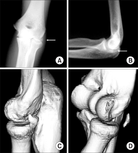
Fig. 1 (A, B) The preoperative anteroposterior and lateral simple radiographs show a radio-opaque density around the radio-capitellar joint (white arrow). (C, D) The reconstructed 3-dimensional computed tomographic images reveal a bony fragment on the posterolateral side of the elbow joint.
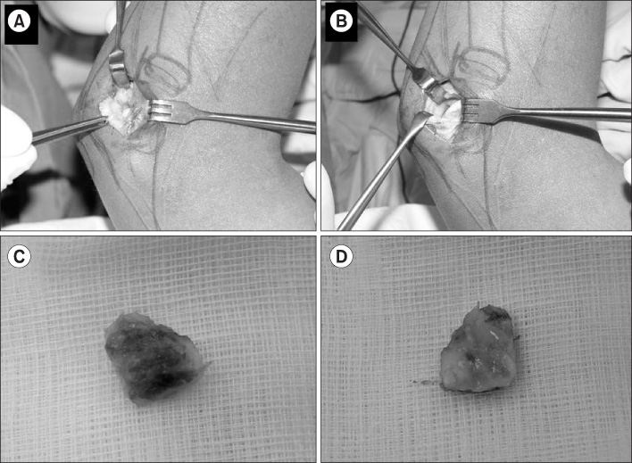
Fig. 2 The operative findings. There was no bruise or evidence of intra-articular bleeding. The mass was encapsulated by the synovium (A) and the surrounding bony structures were intact, including the radial head and capitellum (B). The external surface of the lesion was covered by the joint capsule (C) and the inner surface was covered by the synovial and cartilaginous tissues (D).
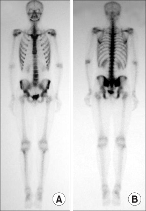
Fig. 3 The bone scintigraphy taken 3 days after excision of the lesion. The absence of a hot spot on the anterior (A) and posterior (B) view implies that there is no acute fracture.

Fig. 4 (A, B) The histopathological findings. The osteochondroid tissue was surrounded by dense fibrous tissue without any evidence of acute hemorrhage (hematoxylin and eosin, ×40 for Fig. 4A and ×100 for Fig. 4B).
Reference
-
1. Bell MS. Loose bodies in the elbow. Br J Surg. 1975. 62:921–924.
Article2. Fahmy NR, Noble J. Ulnar nerve palsy as a complication of synovial osteochondromatosis of the elbow. Hand. 1981. 13:308–310.
Article3. Henderson MS. Loose bodies in the elbow joint. J Am Med Assoc. 1918. xxi:177–180.
Article4. Jeffreys TE. Synovial chondromatosis. J Bone Joint Surg Br. 1967. 49:530–534.
Article5. Jones JR, Evans DM, Kaushik A. Synovial chondromatosis presenting with peripheral nerve compression--a report of two cases. J Hand Surg Br. 1987. 12:25–27.6. Kamineni S, O'Driscoll SW, Morrey BF. Synovial osteochondromatosis of the elbow. J Bone Joint Surg Br. 2002. 84:961–966.
Article7. Matsumoto K, Hukuda S, Fujita M, Kakimoto A, Tachibana S. Cubital bursitis caused by localized synovial chondromatosis of the elbow. A case report. J Bone Joint Surg Am. 1996. 78:275–277.
Article8. Maurice H, Crone M, Watt I. Synovial chondromatosis. J Bone Joint Surg Br. 1988. 70:807–811.
Article9. Milgram JW. Synovial osteochondromatosis: a histopathological study of thirty cases. J Bone Joint Surg Am. 1977. 59:792–801.10. Ruth RM, Groves RJ. Synovial Osteochondromatosis of the elbow presenting with ulnar nerve neuropathy. Am J Orthop. 1996. 25:843–844.
- Full Text Links
-
- Actions
-
Cited
- CITED
-
- Close
- Share
-
- Similar articles
-
- A clinical Study of Synovial Chondromatosis
- Synovial Chondromatosis of the Ulnocarpal Joint
- Synovial Chondromatosis
- Synovial Chondromatosis of the Metacarpophalangeal Joint: A Case Report
- Pigmented Villonodular Synovitis Mimiking the Bone Tumor of the Fossa Olecrani of Elbow in a 8-year-old Boy: A Case Report
