Aberrant Internal Carotid Artery in the Middle Ear
- Affiliations
-
- 1Department of Radiology, Seoul Veterans Hospital, Seoul, Korea. knroo@hanmail.net
- KMID: 2098000
- DOI: http://doi.org/10.3348/jksr.2014.71.4.169
Abstract
- The knowledge about the aberrant internal carotid artery (ICA) in the middle ear is essential for clinicians, because a misdiagnosis of the aberrant ICA could have serious consequences such as excessive aural bleeding during a middle ear surgery. A 38-year-old woman presented with tinnitus and hearing difficulties of the left ear that had started 5 years ago. During otoscopy, an anteroinferior bluish mass was seen in the tympanic space. Computed tomography and magnetic resonance imaging demonstrated a left-side aberrant ICA with bony dehiscence of the carotid canal in the middle ear and a reduced diameter of the tympanic ICA. Herein we report a case of an aberrant ICA in the middle ear. We also review the literature regarding this important vascular anomaly of the temporal bone which may lead to disastrous surgical complications.
MeSH Terms
Figure
-
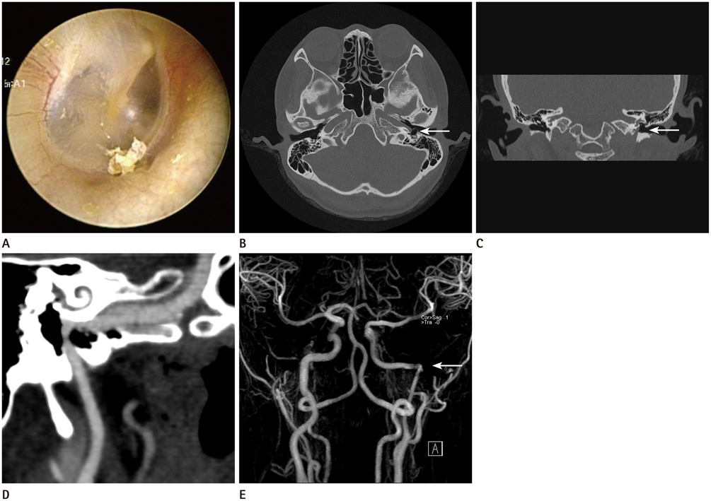
Fig. 1 38-year-old female with tinnitus and hearing difficulty of the left ear history. A. At otoscopy, an anteroinferior bluish mass in the left tympanic space is seen. B. Axial high resolution temporal bone CT scan shows the aberrant left ICA (white arrow), entering the tympanic cavity through a dehiscent carotid plate. C. The left ICA (white arrow) is seen entering the tympanic cavity through the markedly enlarged inferior tympanic canaliculus (Jacobson canal) on coronal temporal bone CT. D. Curved multiplanar reformation CT carotid angiography shows the aberrant left ICA, crossing the cochlear promontory and projected into the tympanic space. E. MRA shows a reduced diameter of the left tympanic ICA (white arrow). The vertical segment of the left ICA is lateral to a line drawn vertically through the vestibule. The aplasia of the A1 segment of the left ACA is revealed. Note.-ACA = anterior cerebral artery, ICA = internal carotid artery, MRA = magnetic resonance angiography
Reference
-
1. Shimizu S, Sasahara G, Iida Y, Shibuya M, Numata T. Aberrant internal carotid artery in the middle ear with a deficiency in the origin of the anterior cerebral artery: a case report. Auris Nasus Larynx. 2009; 36:359–362.2. Sauvaget E, Paris J, Kici S, Kania R, Guichard JP, Chapot R, et al. Aberrant internal carotid artery in the temporal bone: imaging findings and management. Arch Otolaryngol Head Neck Surg. 2006; 132:86–91.3. Eryilmaz A, Dagli M, Cayonu M, Dursun E, Gocer C. An aberrant internal carotid artery in the temporal bone presenting as a middle-ear mass: a case report. J Laryngol Otol. 2008; 122:983–985.4. Lasjaunias P, Santoyo-Vazquez A. Segmental agenesis of the internal carotid artery: angiographic aspects with embryological discussion. Anat Clin. 1984; 6:133–141.5. Koizuka I, Hattori K, Tsutsumi K, Sakuma A, Katsumi N, Kikuchi H, et al. Objective tinnitus caused by an aberrant internal carotid artery. Auris Nasus Larynx. 1998; 25:323–327.6. Botma M, Kell RA, Bhattacharya J, Crowther JA. Aberrant internal carotid artery in the middle-ear space. J Laryngol Otol. 2000; 114:784–787.7. Cole RD, May JS. Aberrant internal carotid artery. South Med J. 1994; 87:1277–1280.8. Duclos JY, Darrouzet V, Martel J, Berge J, Calas V, Bébéar JP. [Abnormal trajectory of the internal carotid artery in the middle ear. Report of a case]. Rev Laryngol Otol Rhinol (Bord). 2000; 121:187–192.9. Ruggles RL, Reed RC. Treatment of aberrant carotid arteries in the middle ear: a report of two cases. Laryngoscope. 1972; 82:1199–1205.
- Full Text Links
-
- Actions
-
Cited
- CITED
-
- Close
- Share
-
- Similar articles
-
- Aberrant Internal Carotid Artery in the Middle Ear: A Case Report
- Aberrant Internal Carotid Artery in the Middle Ear
- One Case of Persistent Stapedial Artery Combined with Aberrant Internal Carotid Artery
- Pulsatile Tinnitus Arising from Aberrant Internal Carotid Artery at Nasopharynx
- Pseudoaneurysm of the Petrosal Internal Carotid Artery in the Middle Ear as a Complication of Middle Ear Cholesteatoma
