J Korean Soc Spine Surg.
2012 Mar;19(1):16-19. 10.4184/jkss.2012.19.1.16.
The Inferior Accessory Ossicle of the Anterior Arch of the Atlas Misdiagnosed as Anterior Arch Fracture: A Case Report
- Affiliations
-
- 1Department of Orthopedic Surgery, National Health Insurance Corporation Ilsan Hospital, Gyeonggi, Korea. esshappy@daum.net
- 2Department of Orthopedic Surgery, Yonsei University College of Medicine, Seoul, Korea.
- KMID: 2097860
- DOI: http://doi.org/10.4184/jkss.2012.19.1.16
Abstract
- STUDY DESIGN: Case report.
OBJECTIVES
We report a very rare case of the inferior accessory ossicle of the anterior arch of the atlas misdiagnosed as anterior arch fracture. SUMMARY OF LITERATURE REVIEW: It is necessary to know the existence of inferior accessory ossicle of the anterior arch of the atlas, even though it is extremely rare.
MATERIALS AND METHODS
A 29-year-old woman was referred to our emergency service unit with symptoms of neck pain and scalp laceration, after being involved in a car accident. She was diagnosed as the inferior accessory ossicle of the anterior arch of the atlas, by multiple diagnostic mordalities.
RESULTS
The symptom of neck pain was relieved spontaneously, and her symptom has been relieved at her latest visit, as a follow up within 3 months.
CONCLUSIONS
It is important to be aware of cervical anatomical variants because we commonly confuse it with other pathologic conditions, such as a fracture and thus, misdiagnose the condition.
Figure
-
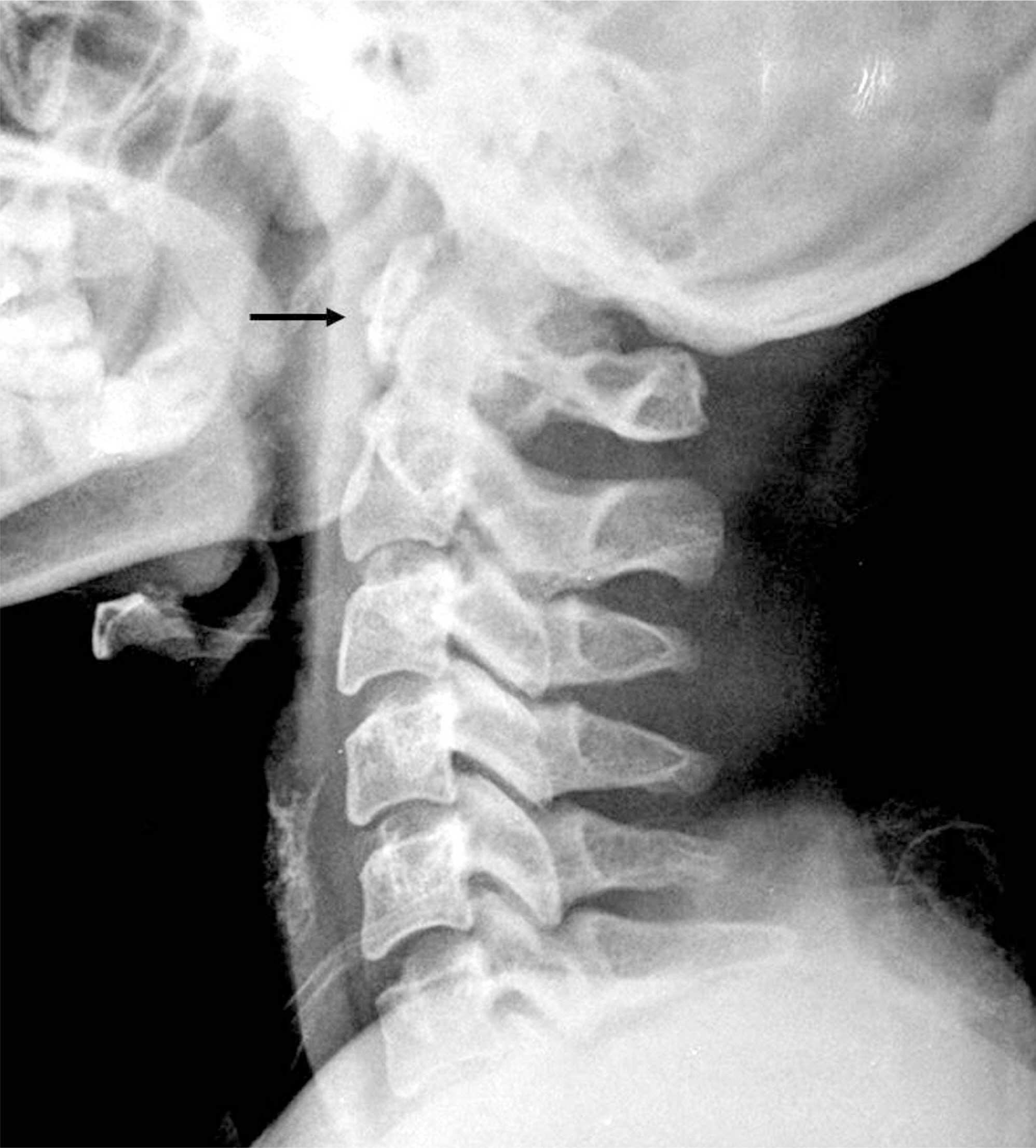
Fig.1. Lateral radiograph of cervical spine shows a well corticated bone fragment inferior to the anterior arch of atlas (black arrow)
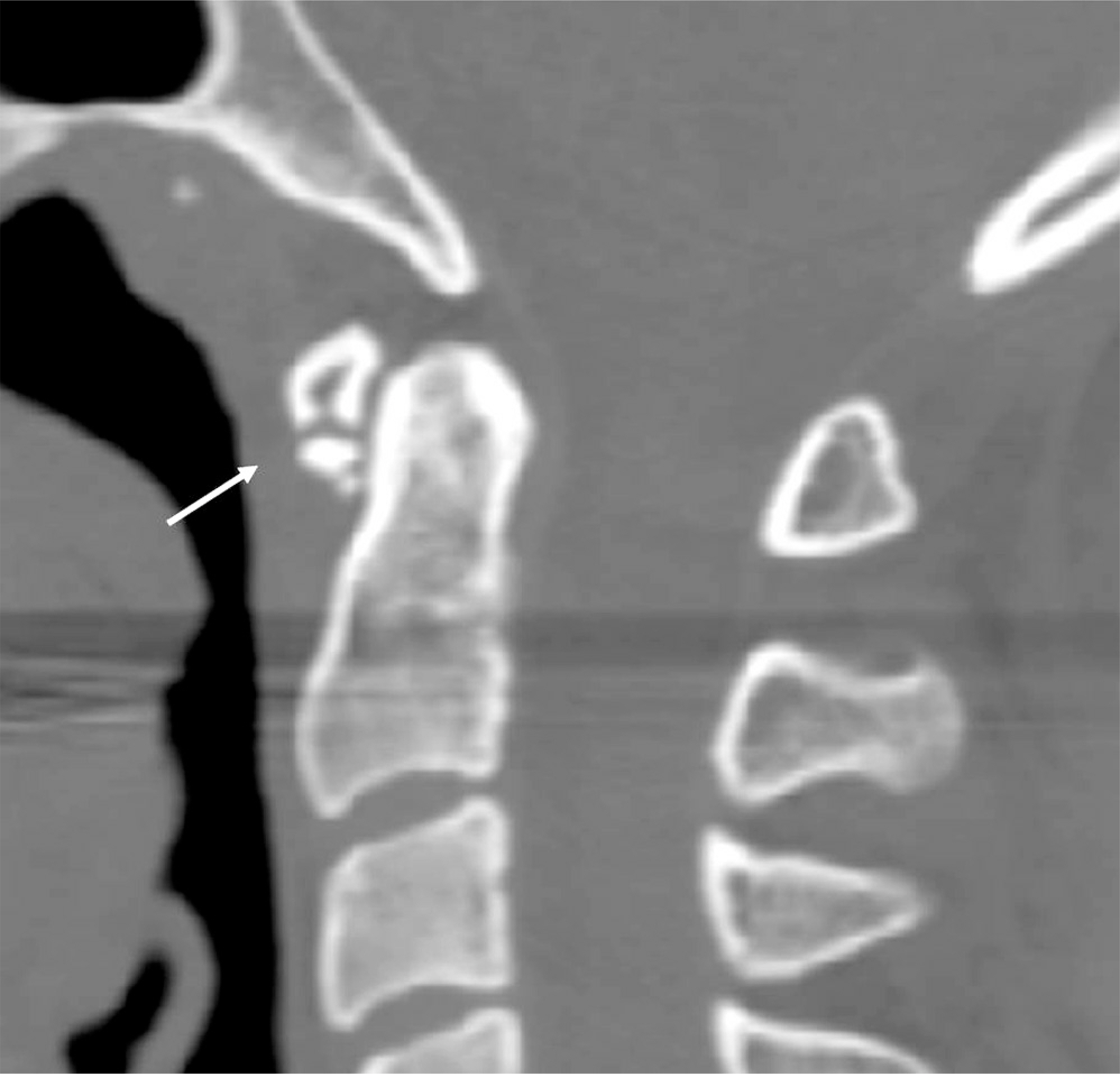
Fig.2. Lateral CT at the level of C1 showing inferior accessory ossicle of the anterior arch of atlas (white arrow).
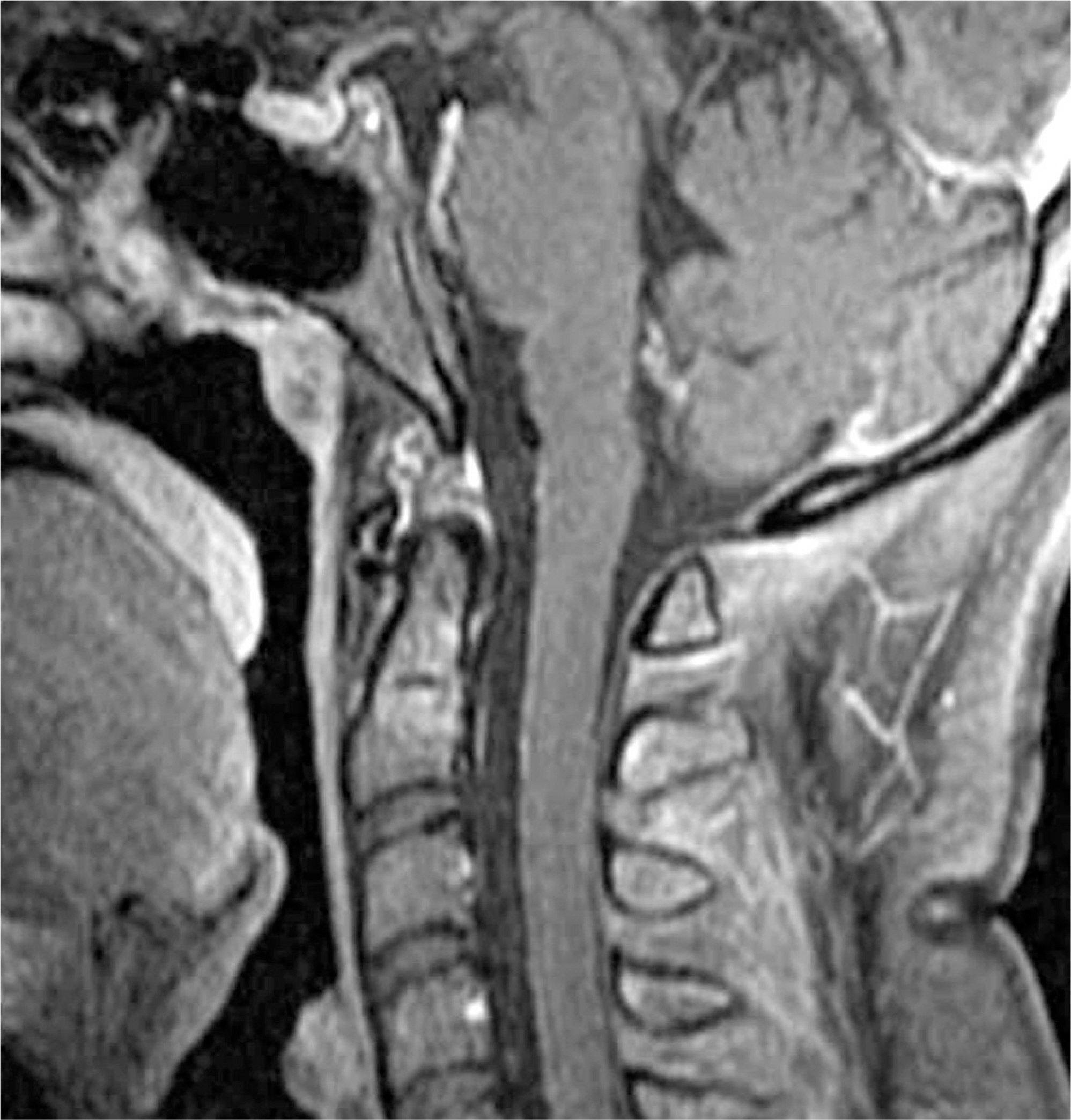
Fig.3. T1 GD enhance MR image showing no definite evidence of hema-toma and soft tissue swelling at the level of C1
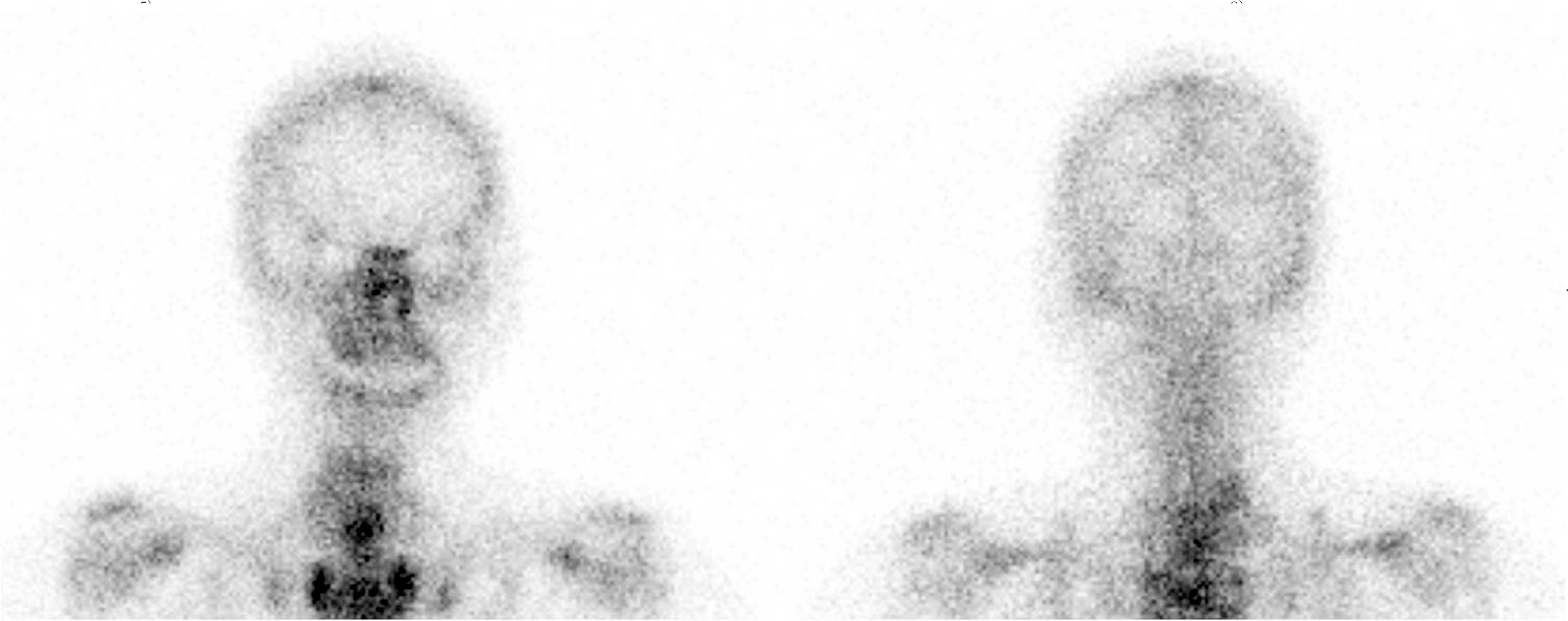
Fig.4. Whole body bone scan showing normal finding.
Reference
-
1.Naji MF., Bhat R. The typical appearance of the inferior accessory ossicle of the anterior arch of the atlas. Surg Radiol Anst. 2009. 31:69–71.
Article2.Kohler A., Zimmer EA. Borderlands of the normal and early pathologic in skeletal roentgenology. Tenth edition. Grune & Stratton Inc., New York,. 1956. 85:206.3.Keats TE. The inferior accessory ossicle of the anterior arch of the atlas. Am J Roentgenol Radium Ther Nucl Med. 1967. 101:834–6.
Article4.Chow K., Motamedi K., Seeger LL., Kalantari BN. Accessory ossicles and sesamoid bones: spectrum of pathology and imaging evaluation. Appl Radiol. 2007. 36:28–37.5.Kim NH., Choi CH., Koh GH. Acute Fractures and Dislocations of the Cervical Spine in Children and Adolescents. J Korean Soc Spine Surg. 1994. 1:19–27.6.Lustrin ES., Karakas SP., Ortiz AO, et al. Pediatric cervical spine: normal anatomy, variants, and trauma. Radiographics. 2003. 23:539–60.
Article7.Von Ludinghausen M., Fahr M., Prescher A, et al. Accessory joints between basiocciput and atlas/axis in the median plane. Clin Anat. 2005. 18:558–71.8.Omezzine SJ., Hafsa C., Lahmar I, et al. Calcific tendinitis of the longus colli: diagnosis by CT. Joint Bone Spine. 2008. 75:90–1.
Article9.Feldman VB. Eagle's syndrome: a case of symptomatic calcification of the styloid ligaments. J Can Chiropr Assoc. 2003. 47:21–7.10.Jevtich V. Horizontal fracture of the anterior arch of the atlas. Case report. J Bone Joint Surg Am. 1986. 68:1094–5.
Article
- Full Text Links
-
- Actions
-
Cited
- CITED
-
- Close
- Share
-
- Similar articles
-
- Inferior Accessory Ossicle of the Anterior Arch of the Atlas
- Stress Fracture of the Anterior Atlas Arch Following C1 Posterior Arch Resection for Cervical Myelopathy with Retro-Odontoid Pseudotumor
- Spontaneous Anterior Atlas Fracture Following C1 Laminectomy without Fusion: A Case Report
- Congenital Anomaly of the Atlas Misdiagnosed as Posterior Arch Fracture of the Atlas and Atlantoaxial Subluxation
- Congenital Hypoplasia of the Posterior Arch of the Atlas Associated with a Fracture of the Odontoid Process: A Case Report
