Pseudomeningocele After Spine Surgery: 3 cases of different symptoms
- Affiliations
-
- 1Department of Orthopedic Surgery, Seoul Sacred Heart General Hospital, Seoul, Korea. adkajs@hanmail.net
- KMID: 2097830
- DOI: http://doi.org/10.4184/jkss.2006.13.2.132
Abstract
- Pseudomeningocele after spine surgery can cause various symptoms, but it can also be silent. We experienced 3 cases of pseudomeningocele with different symptoms and we analyzed the characteristics of each case. A small pseudomeningocele without connection to the subarachnoidal space can show no symptoms. A pseudomeningocele with a small dural tear and it's abutted on the duramater at a small portion can produce sciatica and limitations of straight leg raising due to adhesion of the cauda equina around the dural tear. In addition, a large pseudomeningocele with a big dural and lamina defect can produce back tenderness furthermore, a patient with such a lesion can have low back pain and leg pain that are aggravated by an increment of abdominal pressure or by impact to the body and even by walking. Pseudomeningocele should be suspected when symptoms recur after spine surgery and especially in the case of dural tear during an operation
Keyword
Figure
-

Fig. 1. These are images of 55 year old female patient who underwent multiple schwanommas excision at T11, L3 and PLIF L4-5 for spondylolytic spondylolisthesis. (A) Sagittal MR T2 weighted image which shows large well marginated cyst in size of 160 50 mm connected to subarachnoid space(arrow). (B) Intraoperative finding shows dural defect. Through the defect, inflamed cauda equina are seen and some of them are adhesed to the margin. (C) Intraoperative finding shows fascia lata graft covering the dural defect. (D) Sagittal MR T2 weighted image at 24 months after dural repair which shows complete obliteration of the cyst.

Fig. 2. This is a sagittal MR T2 weighted image of 60 year old female patient who underwent PLF L4-5 which shows 60 25 mm sized cyst that seems to be connected to subarachnoid space.
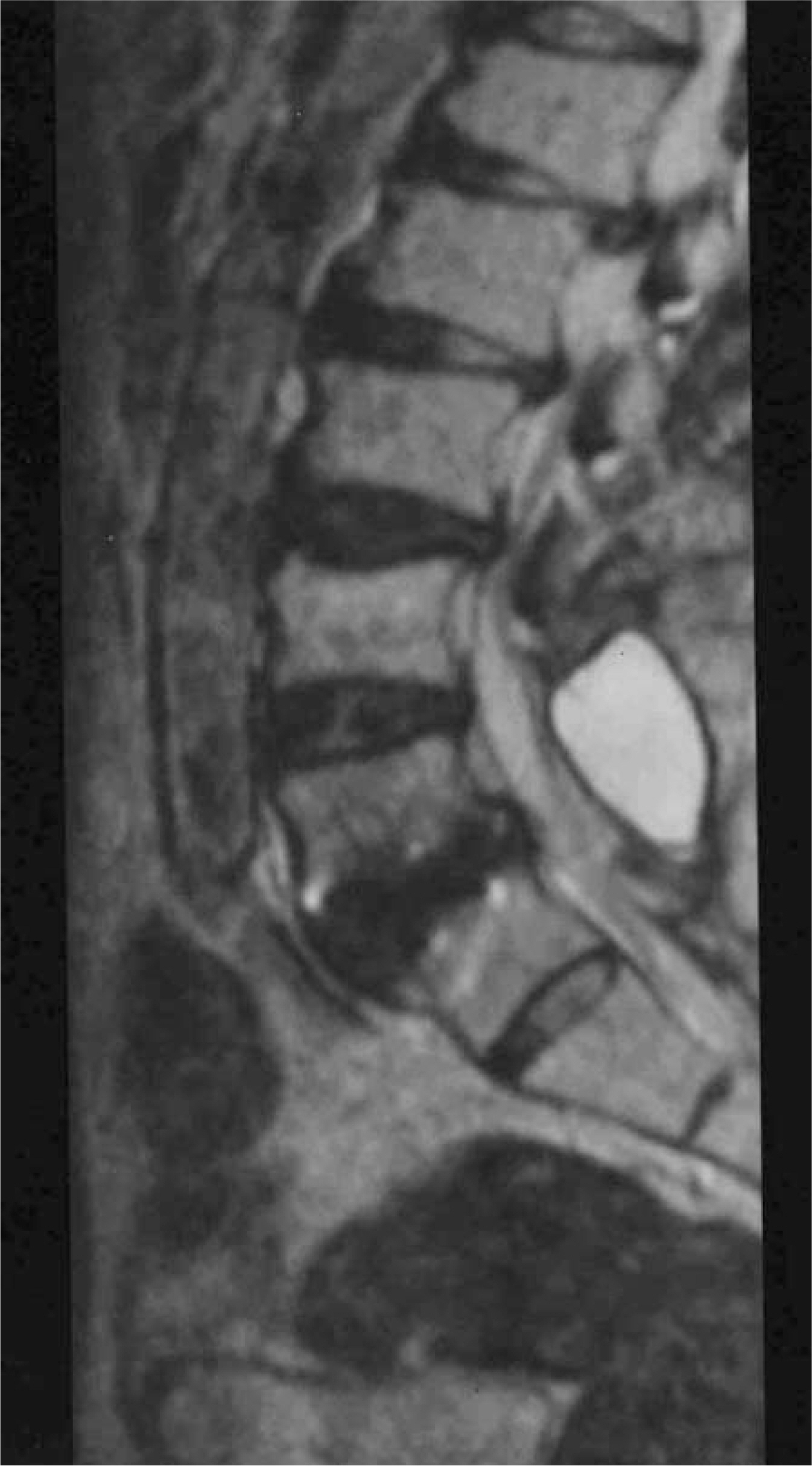
Fig. 3. This is a sagittal MR T2 weighted image of 67 year old male patient who underwent PLF L3-4-5 which shows 40 25 mm sized cyst that seems not to be connected to subarachnoid space.
Reference
-
01). Lee KS., Hardy IM. Postlaminectomy lumbar pseudomeningocele: report of four cases. J Neurogurg. 1992. 30:111–114.02). McCormack BM., Tayler SL., Health S., Scanlon J. Pseudomeningocele/CSF fistula in a patient with lumbar spinal implants treated with epidural blood patch and a brief course of closed subarachnoid drainage: A case report. Spine. 1996. 21:2273–2276.03). O' Connor D., Maskery N., Griffiths WE. Pseudomeningocele nerve root entrapment after lumbar discectomy. Spine. 1998. 23:1501–1502.04). Rinaldi I., Hodges TO. Iatrogenic lumbar meningocele: report of three cases. J Neurol Neurosurg Psychiat. 1970. 33:484–492.05). Panolini S., Ciappetta P., Piattella MC. Intraspinous postlaminectomy pseudomeningocele. Eur Spine J. 2003. 12:325–327.
Article06). Hyndman OR., Gerber WF. Spinal extradural cysts, con-genital and acquired. Report of cases. J Neurosurg. 1946. 3:474–486. (cited from Rinaldi I, Hodges TO: Iatrogenic lumbar meningocele: report of three cases. J Neurol Neurosurg Psychiat 1970; 33: 484-492.).07). Swanson HS., Fincher EF. Extradural arachnoidal cyst of traumatic origin. J Neurosurg. 1947. 4:530–538. (cited from Rinaldi I, Hodges TO: Iatrogenic lumbar meningocele: report of three cases. J Neurol Neurosurg Psychiat 1970; 33: 484-492.).08). Teplick JG., Teplick SK., Goodman LR., Haskin ME. Pitfalls and unusual findings in computed tomography of the lumbar spine. J Comput Assist Tomogr. 1982. 6:888–893.
Article09). Aldrete AJ., Ghaly R. Postlaminectomy pseudomeningocele: An unsuspected cause of low back pain. Regional Anesthesia. 1995. 20:75–79.10). Vinas F., Slade H. Meningocele as complication of laminectomy. Rev Med Cordoba. 1959. 47:470–473. (cited from Rinaldi I, Hodges TO: Iatrogenic lumbar meningo-cele: report of three cases. J Neurol Neurosurg Psychiat 1970; 33: 484-492.).11). Misra SN., Morgan HW., Sedler R. Lumbar myofascial flap for pseudomeningocele repair. Neurosurg Focus. 2003. 15(3):e13.
Article
- Full Text Links
-
- Actions
-
Cited
- CITED
-
- Close
- Share
-
- Similar articles
-
- Clinical Experience of Pain Management for Postlaminectomy Syndrome due to Pseudomeningocele: A case report
- A Case of Post-Traumatic Pseudomeningocele Treated by Lumboperitoneal Shunt
- Spinal anesthesia in a patient with postoperative iatrogenic pseudomeningocele: A case report
- Iatrogenic Pseudomeningomyelocele after Lumbar Laminectomies: Report of Cases
- Pseudomeningocele after lumbar discectomy treated with fibrin glue and epidural blood patch: A case report
