J Adv Prosthodont.
2015 Oct;7(5):349-357. 10.4047/jap.2015.7.5.349.
Effects of core characters and veneering technique on biaxial flexural strength in porcelain fused to metal and porcelain veneered zirconia
- Affiliations
-
- 1Department of Prosthodontics, School of Dentistry and Institute of Oral Bio-Science, Chonbuk National University, Jeonju, Republic of Korea. jmseo@jbnu.ac.kr
- 2Biomedical Research Institute, Chonbuk National University Hospital, Jeonju, Republic of Korea.
- 3Department Dental Biomaterials, Institute of Oral Bio-science, School of Dentistry, Chonbuk National University, Jeonju, Republic of Korea.
- KMID: 2083877
- DOI: http://doi.org/10.4047/jap.2015.7.5.349
Abstract
- PURPOSE
The purpose of this study was to assess the impact of the core materials, thickness and fabrication methods of veneering porcelain on prosthesis fracture in the porcelain fused to metal and the porcelain veneered zirconia.
MATERIALS AND METHODS
Forty nickel-chrome alloy cores and 40 zirconia cores were made. Half of each core group was 0.5 mm-in thickness and the other half was 1.0 mm-in thickness. Thus, there were four groups with 20 cores/group. Each group was divided into two subgroups with two different veneering methods (conventional powder/liquid layering technique and the heat-pressing technique). Tensile strength was measured using the biaxial flexural strength test based on the ISO standard 6872:2008 and Weibull analysis was conducted. Factors influencing fracture strength were analyzed through three-way ANOVA (alpha< or =.05) and the influence of core thickness and veneering method in each core materials was assessed using two-way ANOVA (alpha< or =.05).
RESULTS
The biaxial flexural strength test showed that the fabrication method of veneering porcelain has the largest impact on the fracture strength followed by the core thickness and the core material. In the metal groups, both the core thickness and the fabrication method of the veneering porcelain significantly influenced on the fracture strength, while only the fabrication method affected the fracture strength in the zirconia groups.
CONCLUSION
The fabrication method is more influential to the strength of a prosthesis compared to the core character determined by material and thickness of the core.
Figure
-
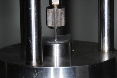
Fig. 1 Biaxial flexural strength was measured by the universal testing machine.
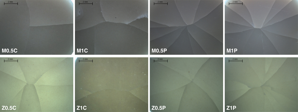
Fig. 2 Fracture status of the fragmented sample was observed through the optical microscope.
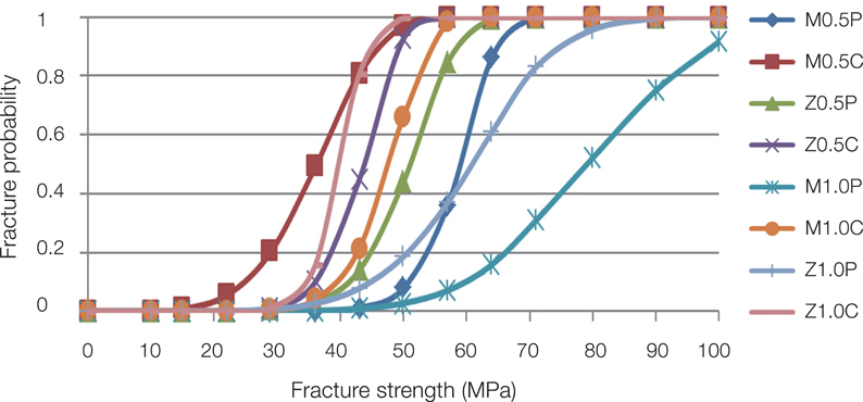
Fig. 3 Probability Weibull was analyzed for group comparisons.
Reference
-
1. McLean JW, Hughes TH. The reinforcement of dental porcelain with ceramic oxides. Br Dent J. 1965; 119:251–267.2. Magne P, Belser U. The esthetic width in fixed porcelain restorations. J Prosthodont. 1999; 8:106–118.3. Guazzato M, Albakry M, Ringer SP, Swain MV. Strength, fracture toughness and microstructure of a selection of allceramic materials. Part II. Zirconia-based dental ceramics. Dent Mater. 2004; 20:449–456.4. Denry I, Kelly JR. State of the art of zirconia for dental applications. Dent Mater. 2008; 24:299–307.5. Triwatana P, Nagaviroj N, Tulapornchai C. Clinical performance and failures of zirconia-based fixed partial dentures: a review literature. J Adv Prosthodont. 2012; 4:76–83.6. Näpänkangas R, Raustia A. Twenty-year follow-up of metalceramic single crowns: a retrospective study. Int J Prosthodont. 2008; 21:307–311.7. Reitemeier B, Hänsel K, Kastner C, Walter MH. Metal-ceramic failure in noble metal crowns: 7-year results of a prospective clinical trial in private practices. Int J Prosthodont. 2006; 19:397–399.8. Sailer I, Fehér A, Filser F, Gauckler LJ, Lüthy H, Hämmerle CHF. Five-year clinical results of zirconia frameworks for posterior fixed partial dentures. Int J Prosthodont. 2007; 20:383–388.9. Sailer I, Fehér A, Filser F, Lüthy H, Gauckler LJ, Scharer P, Hämmerle CHF. Prospective clinical study of zirconia posterior fixed partial dentures: 3-year follow-up. Quintessence Int. 2006; 37:685–693.10. Agustín-Panadero R, Román-Rodríguez JL, Ferreiroa A, Solá-Ruíz MF, Fons-Font A. Zirconia in fixed prosthesis: A literature review. J Clin Exp Dent. 2014; 6:e66–e73.11. Quinn JB, Sundar V, Parry EE, Quinn GD. Comparison of edge chipping resistance of PFM and veneered zirconia specimens. Dent Mater. 2010; 26:13–20.12. Alhasanyah A, Vaidyanathan TK, Flinton RJ. Effect of core thickness differences on post-fatigue indentation fracture resistance of veneered zirconia crowns. J Prosthodont. 2013; 22:383–390.13. Millen CS, Reuben RL, Ibbetson RJ. The effect of coping/ veneer thickness on the fracture toughness and residual stress of implant supported, cement retained zirconia and metal-ceramic crowns. Dent Mater. 2012; 28:e250–e258.14. Henriques B, Soares D, Silva FS. Shear bond strength of a hot pressed Au-Pd-Pt alloy-porcelain dental composite. J Mech Behav Biomed Mater. 2011; 4:1718–1726.15. Rice RW. Limitations of pore-stress concentrations on the mechanical properties of porous materials. J Mater Sci. 1997; 32:4731–4736.16. Anusavice KJ, Phillips RW. Science of dental materials. 11th ed. St. Louis, Mo.: Saunders;2003. p. 665–719.17. Nakamura T, Wakabayashi K, Kawamura Y, Kinuta S, Mutobe Y, Yatani H. Analysis of internal defects in all-ceramic crowns using micro-focus X-ray computed tomography. Dent Mater J. 2007; 26:598–601.18. Ban S, Anusavice KJ. Influence of test method on failure stress of brittle dental materials. J Dent Res. 1990; 69:1791–1799.19. Ban S, Hasegawa J, Anusavice KJ. Effect of loading conditions on bi-axial flexure strength of dental cements. Dent Mater. 1992; 8:100–104.20. Lin WS, Ercoli C, Feng C, Morton D. The effect of core material, veneering porcelain, and fabrication technique on the biaxial flexural strength and weibull analysis of selected dental ceramics. J Prosthodont. 2012; 21:353–362.21. Aboushelib MN, de Kler M, van der Zel JM, Feilzer AJ. Effect of veneering method on the fracture and bond strength of bilayered zirconia restorations. Int J Prosthodont. 2008; 21:237–240.22. Aboushelib MN, Kleverlaan CJ, Feilzer AJ. Microtensile bond strength of different components of core veneered all-ceramic restorations. Part II: Zirconia veneering ceramics. Dent Mater. 2006; 22:857–863.23. Aboushelib MN, Kleverlaan CJ, Feilzer AJ. Microtensile bond strength of different components of core veneered all-ceramic restorations. Part 3: double veneer technique. J Prosthodont. 2008; 17:9–13.24. Christensen GJ. PFM vs zirconia restorations-how are they comparing clinically? Clinician's Report. CR found. 2008; 11:1–2.25. Gonzaga CC, Cesar PF, Miranda WG Jr, Yoshimura HN. Slow crack growth and reliability of dental ceramics. Dent Mater. 2011; 27:394–406.
- Full Text Links
-
- Actions
-
Cited
- CITED
-
- Close
- Share
-
- Similar articles
-
- Shear bond strength of veneering porcelain to zirconia and metal cores
- COMPARISON OF COLOR AND OPACITY OF COPY-MILLED IN-CREAM ALUMINA CORE AND SPINELL CORE
- A study on the shear bond strengths of veneering ceramics to the colored zirconia core
- The effect of surface finishes on flexural strength, fracture toughness of feldspathic dental porcelain
- The effect of zirconia surface architecturing technique on the zirconia/veneer interfacial bond strength
