Korean J Hematol.
2005 Dec;40(4):278-281. 10.5045/kjh.2005.40.4.278.
Acute Torsion of a Wandering Spleen Managed by Splenopexy
- Affiliations
-
- 1Department of Pediatrics, Chungnam National University College of Medicine, Daejeon, Korea. sunyoung@cnuh.co.kr
- 2Department of Surgery, Chungnam National University College of Medicine, Daejeon, Korea.
- KMID: 2083468
- DOI: http://doi.org/10.5045/kjh.2005.40.4.278
Abstract
- Torsion of the spleen is a rare cause of abdominal pain in children and it may occur in conjunction with wandering spleen. Wandering spleen is the presence of the spleen in a location other than the left upper quadrant, and it is secondary to the congenital or functional absence of splenic ligaments. The occurrence of wandering spleen is rare in adults and it's even less common in children. The most common presentation is acute abdominal pain, although the signs and symptoms vary widely. Due to the risk of splenic infarction, making a rapid and accurate diagnosis is essential. When a wandering spleen is diagnosed, the treatment of choice is splenopexy, even if the patient is asymptomatic. If splenic necrosis is present, then splenectomy is usually required. We describe here a 4-year-old girl with torsion of a wandering spleen that was managed by splenopexy.
Keyword
MeSH Terms
Figure
-
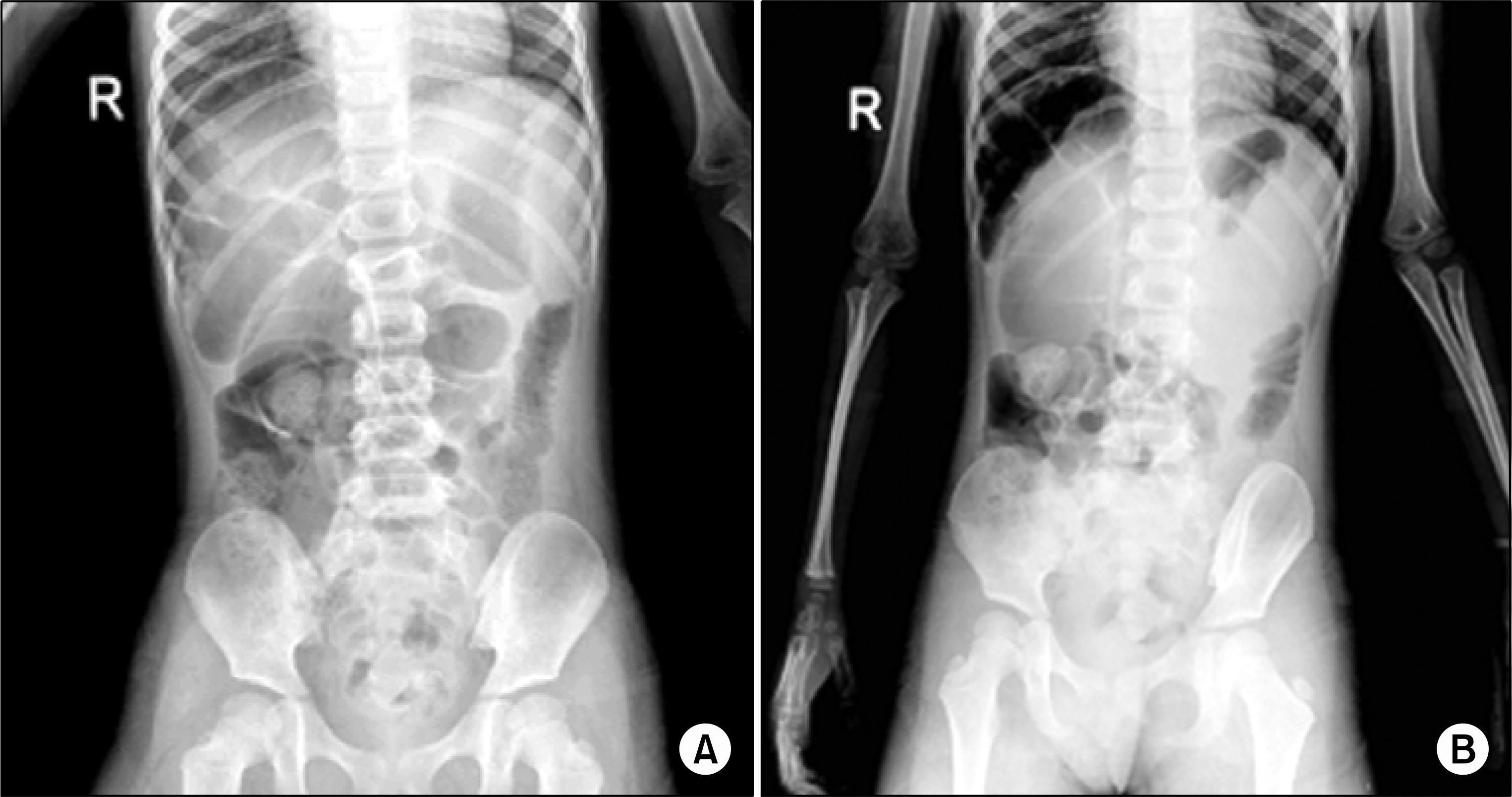
Fig. 1. (A) Normal spleen shadow is shown in supine position. (B) But at erect position, spleen shadow is shown in the lower-medial of normal position.
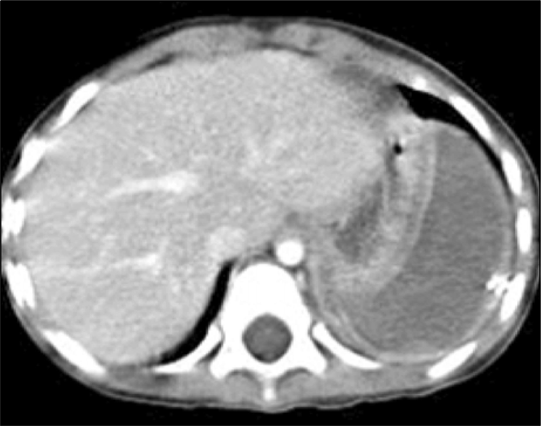
Fig. 2. Abdominal CT. Hypoperfusion of enlarged spleen on contrast enhanced scan, suggesting splenic infarction.
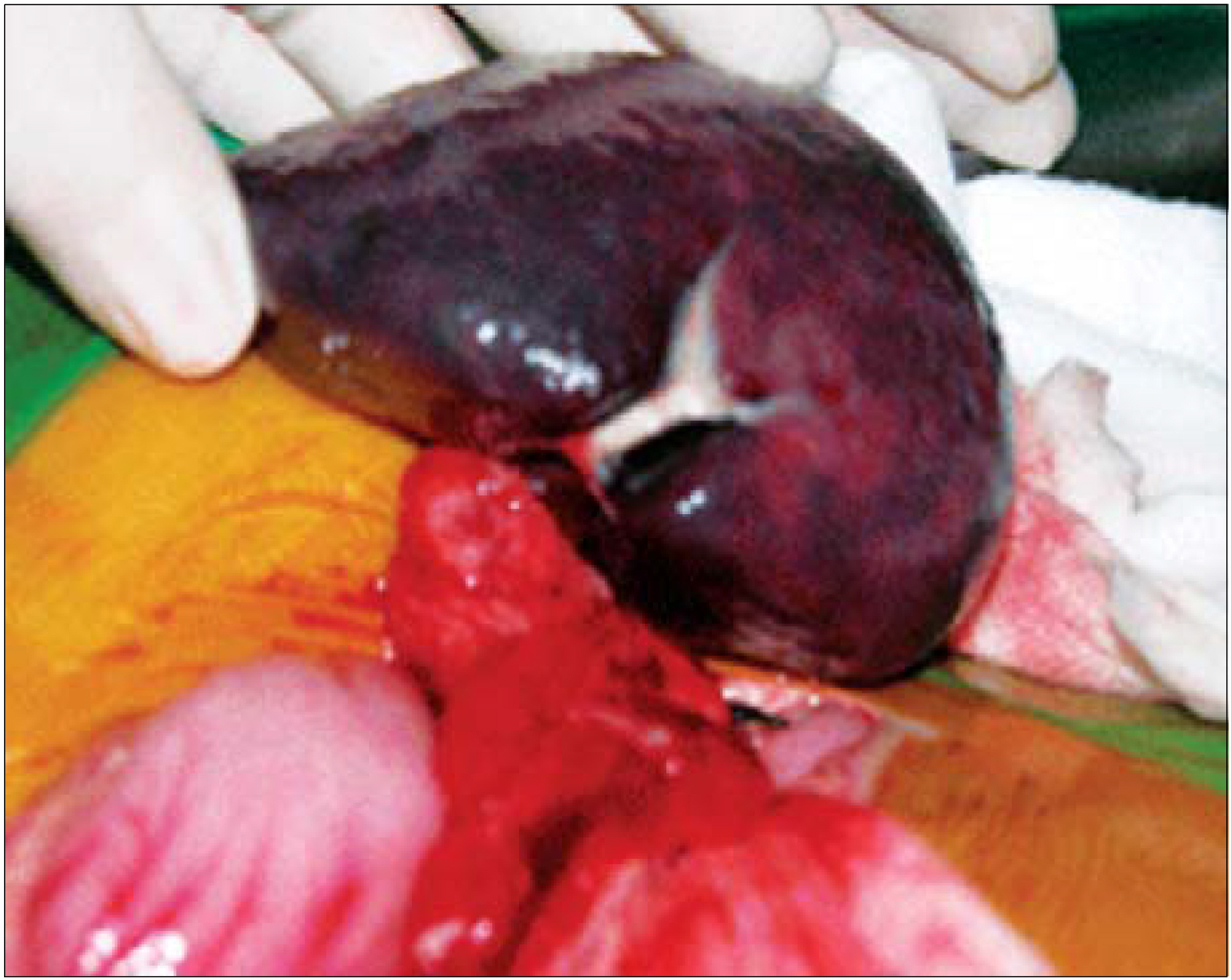
Fig. 3. Ischemic change of the spleen has taken place. Mobile wandering spleen with no attachment except the main vascular pedicle. 360 degree torsion of splenic hilum is shown, and also inflammation of surrounding tissues.
Reference
-
1). Romero JR, Barksdale EM Jr. Wandering spleen: a rare cause of abdominal pain. Pediatr Emerg Care. 2003; 19:412–4.
Article2). Brown CV, Virgilio GR, Vazquez WD. Wandering spleen and its complications in children: a case series and review of the literature. J Pediatr Surg. 2003; 38:1676–9.
Article3). Cavazos S, Ratzer ER, Fenoglio ME. Laparoscopic management of the wandering spleen. Laparoen dosc Adv Surg Tech A. 2004; 14:227–9.
Article4). Eraklis AJ, Filler RM. Splenectomy in childhood: a review of 1413 cases. J Pediatr Surg. 1972; 7:382–8.
Article5). Buehner M, Baker MS. The wandering spleen. Surg Gynecol Obstet. 1992; 175:373–87.6). Rodkey ML, Macknin ML. Pediatric wandering spleen: case report and review of the literature. Clin Pediatr. 1992; 31:289–94.7). Heydenrych JJ, Du Toit DF. Torsion of the spleen and associated ‘rune belly syndrome’. A case report and review of the literature. S Afr Med J. 1978; 53:637–9.8). Pearson JB. Torsion of the spleen associated with congenital absence of the left kidney. Br J Surg. 1964; 51:393–5.
Article9). Spector JM, Chappell J. Gastric volvulus associated with wandering spleen in a child. J Pediatr Surg. 2000; 35:641–2.
Article10). Gordon DH, Burreli MI, Levin DC, Mueller CF, Becker JA. Wandering spleen the radiological and cllinical spectrum. Radiology. 1977; 125:39–46.11). Allen KB, Andrews G. Pediatric wandering spleen the case for splenopexy: review of 35 reported cases in the literature. J Pediatr Surg. 1989; 24:432–5.12). Thompson JS, Ross RJ, Pizzaro ST. The wandering spleen in infancy and childhood. Clin Pediatr Phila. 1980; 19:221–4.
Article13). Tait NP, Young JR. The wandering spleen: an ultrasonic diagnosis. J Clin Utrasound. 1985; 13:141–4.
Article14). Nemcek AA Jr, Miller FH, Fitzgerald SW. Acute torsion of a wandering spleen: diagnosis by CT and duplex Doppler and color flow sonography. AJR Am J Roentgenol. 1991; 157:307–9.
Article15). Martinez Ferro M, Elmo G, Laje P. Laparoscopic pocket splenopexy for wandering spleen: a case report. J Pediatr Surg. 2005; 40:882–4.
- Full Text Links
-
- Actions
-
Cited
- CITED
-
- Close
- Share
-
- Similar articles
-
- Infarction of Wandering Spleen with Torsion of Its Pedicle: A case report
- Splenopexy for Wandering Spleen with Torsion in a Child
- Torsion of a Wandering Spleen Treated by Laparoscopic Surgery: A Case Report
- A Case of Torsion of Wandering Spleen
- Multidetector Computed Tomography Findings of Mesenteroaxial Gastric Volvulus Combined with Torsion of Wandering Spleen: A Case Report and Literature Review
