Development of an Online Radiology Case Review System Featuring Interactive Navigation of Volumetric Image Datasets Using Advanced Visualization Techniques
- Affiliations
-
- 1Department of Radiology, Seoul National University Bundang Hospital, Seongnam, Korea.
- 2Department of Radiology, SMG-SNU Boramae Medical Center, Seoul, Korea. mdwoo79@gmail.com
- 3Department of Computer Science and Engineering, Seoul National University, Seoul, Korea.
- 4Department of Radiology, Soonchunhyang University Bucheon Hospital, Bucheon, Korea.
- 5Program in Biomedical Radiation Sciences, Department of Transdisciplinary Studies, Graduate School of Convergence Science and Technology, Seoul National University, Suwon, Korea.
- KMID: 2079566
- DOI: http://doi.org/10.3348/jksr.2015.73.5.312
Abstract
- PURPOSE
To develop an online radiology case review system that allows interactive navigation of volumetric image datasets using advanced visualization techniques.
MATERIALS AND METHODS
Our Institutional Review Board approved the use of the patient data and waived the need for informed consent. We determined the following system requirements: volumetric navigation, accessibility, scalability, undemanding case management, trainee encouragement, and simulation of a busy practice. The system comprised a case registry server, client case review program, and commercially available cloud-based image viewing system.
RESULTS
In the pilot test, we used 30 cases of low-dose abdomen computed tomography for the diagnosis of acute appendicitis. In each case, a trainee was required to navigate through the images and submit answers to the case questions. The trainee was then given the correct answers and key images, as well as the image dataset with annotations on the appendix. After evaluation of all cases, the system displayed the diagnostic accuracy and average review time, and the trainee was asked to reassess the failed cases. The pilot system was deployed successfully in a hands-on workshop course.
CONCLUSION
We developed an online radiology case review system that allows interactive navigation of volumetric image datasets using advanced visualization techniques.
MeSH Terms
Figure
-

Fig. 1 Diagram showing overall architecture of the system. The system comprised a case registry server, cloud image server, client case review program, and client case image viewer.
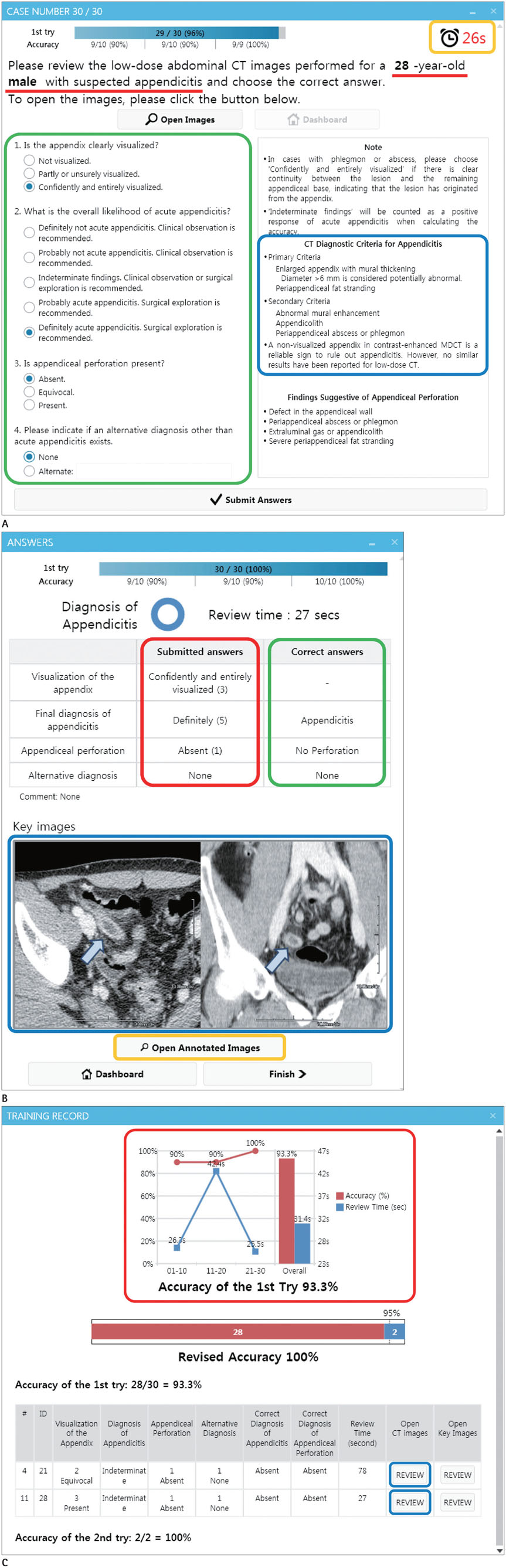
Fig. 2 Screenshots of the client case review program. A. A question page shows brief clinical findings (red underline), case questions (green box), diagnostic criteria for appendicitis (blue box), and the time spent reviewing each case (orange box). B. A feedback page shows the submitted answers (red box), correct answers (green box), and annotated key images (blue box). The trainee can open the annotated image dataset in the client image viewer by clicking the 'Open Annotated Images' button (orange box). C. A training record page shows the trainee's diagnostic accuracy and average review time per case (red box). The trainees can reassess their failed cases by clicking 'Review' buttons (blue box).
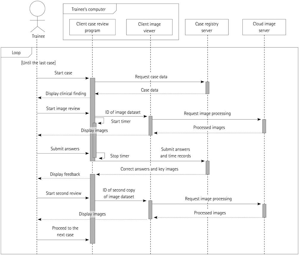
Fig. 3 Sequence diagram showing interactions during a case review cycle. In each case review, the client case review program retrieved the case data, including brief clinical findings, case questions, and the identification number of the image dataset (the first copy without annotations), from the case registry server and displayed the brief clinical findings to the trainee. After receiving the trainee's request, the client case review program sent a command that included the identification number of the image dataset to the client image viewer, and the client image viewer then displayed the processed images received from the cloud image server for the trainee to review. During the review process, the client case review program displayed the time spent reviewing each case. The review time and answers submitted for each case were stored in the case registry server. After the trainee submitted the answers for each case, the client case review program received the correct answers and key images from the case registry server, and displayed them to the trainee as feedback. Following the trainee's request, the client case review program sent a command containing the identification number of the annotated image dataset (the second copy with the annotations for the appendix and other important image findings) to the client image viewer, and the trainee could navigate through the annotated image dataset using the client image viewer. The boxes with dashed vertical lines represent lifelines of the four components of the system and a trainee. The vertical gray bars on the lifelines represent executions. The solid horizontal arrows represent messages between the lifelines and dotted horizontal arrows represent reply messages. ID = identification number
Cited by 1 articles
-
Central Image Archiving and Management System for Multicenter Clinical Studies: Lessons from Low-dose CT for Appendicitis Trial
Yousun Ko, Jin Woo Choi, Dong Hyun Kim, Kyong Joon Lee, Sang Soo Shin, Ji Young Woo, Seong Whi Cho, Bong Soo Kim, Kyoung Ho Lee
J Korean Soc Radiol. 2017;76(3):165-172. doi: 10.3348/jksr.2017.76.3.165.
Reference
-
1. Williams J, Sato TS, Policeni B. Pulmonary embolism teaching file: a simple pilot study for rapidly increasing pulmonary embolism recognition among new residents using interactive cross-sectional imaging. Acad Radiol. 2013; 20:1048–1051.2. Bailey JH, Roth TD, Kohli MD, Heitkamp DE. Real view radiology-impact on search patterns and confidence in radiology education. Acad Radiol. 2014; 21:859–868.3. Rojas CA, Jawad H, Chung JH. The new era of radiology teaching files. AJR Am J Roentgenol. 2012; 198:773–776.4. Phillips GS, LoGerfo SE, Richardson ML, Anzai Y. Interactive Web-based learning module on CT of the temporal bone: anatomy and pathology. Radiographics. 2012; 32:E85–E105.5. Andriole KP, Wolfe JM, Khorasani R, Treves ST, Getty DJ, Jacobson FL, et al. Optimizing analysis, visualization, and navigation of large image data sets: one 5000-section CT scan can ruin your whole day. Radiology. 2011; 259:346–362.6. Bhargavan M, Kaye AH, Forman HP, Sunshine JH. Workload of radiologists in United States in 2006-2007 and trends since 1991-1992. Radiology. 2009; 252:458–467.7. Shaffer K. Radiology education in the digital era. Radiology. 2005; 235:359–360.8. Rowell MR, Johnson PT, Fishman EK. Radiology education in 2005: world wide web practice patterns, perceptions, and preferences of radiologists. Radiographics. 2007; 27:563–571.9. Bandukwala T, Arora S, Athreya S. Net assets: review of online radiology resources. Part I. Educational resources. Radiology. 2011; 261:350–356.10. Bhargava P, Dhand S, Lackey AE, Pandey T, Moshiri M, Jambhekar K. Radiology education 2.0--on the cusp of change: part 2. eBooks; file sharing and synchronization tools websites/teaching files; reference management tools and note taking applications. Acad Radiol. 2013; 20:373–381.11. Korbage AC, Bedi HS. Mobile technology in radiology resident education. J Am Coll Radiol. 2012; 9:426–429.12. Kahn CE Jr, Santos A, Thao C, Rock JJ, Nagy PG, Ehlers KC. A presentation system for just-in-time learning in radiology. J Digit Imaging. 2007; 20:6–16.13. Rowe SP, Siddiqui A, Bonekamp D. The key image and case log application: new radiology software for teaching file creation and case logging that incorporates elements of a social network. Acad Radiol. 2014; 21:916–930.14. Balkman JD, Loehfelm TW. A cloud-based multimodality case file for mobile devices. Radiographics. 2014; 34:863–872.15. Rengier F, Häfner MF, Unterhinninghofen R, Nawrotzki R, Kirsch J, Kauczor HU, et al. Integration of interactive three-dimensional image post-processing software into undergraduate radiology education effectively improves diagnostic skills and visual-spatial ability. Eur J Radiol. 2013; 82:1366–1371.16. Arya R, Morrison T, Zumwalt A, Shaffer K. Making education effective and fun: stations-based approach to teaching radiology and anatomy to third-year medical students. Acad Radiol. 2013; 20:1311–1318.17. Zou L, King A, Soman S, Lischuk A, Schneider B, Walor D, et al. Medical students' preferences in radiology education a comparison between the Socratic and didactic methods utilizing powerpoint features in radiology education. Acad Radiol. 2011; 18:253–256.18. TeraRecon. iNtuition. Accessed July 21, 2014. Available at: http://www.terarecon.com/wordpress/wp-content/uploads/2011/11/iNtuition-2011.pdf.19. Strub WM, Vagal AA, Tomsick T, Moulton JS. Overnight resident preliminary interpretations on CT examinations: should the process continue? Emerg Radiol. 2006; 13:19–23.20. Hunter TB, Taljanovic MS, Krupinski E, Ovitt T, Stubbs AY. Academic radiologists' on-call and late-evening duties. J Am Coll Radiol. 2007; 4:716–719.21. Paulson EK, Kalady MF, Pappas TN. Clinical practice. Suspected appendicitis. N Engl J Med. 2003; 348:236–242.22. Kim K, Kim YH, Kim SY, Kim S, Lee YJ, Kim KP, et al. Low-dose abdominal CT for evaluating suspected appendicitis. N Engl J Med. 2012; 366:1596–1605.23. Kim SY, Lee KH, Kim K, Kim TY, Lee HS, Hwang SS, et al. Acute appendicitis in young adults: low- versus standard-radiation-dose contrast-enhanced abdominal CT for diagnosis. Radiology. 2011; 260:437–445.24. Park JH. LOCAT Group. Diagnostic imaging utilization in cases of acute appendicitis: multi-center experience. J Korean Med Sci. 2014; 29:1308–1316.25. Ahn S. LOCAT group. LOCAT (low-dose computed tomography for appendicitis trial) comparing clinical outcomes following low- vs standard-dose computed tomography as the first-line imaging test in adolescents and young adults with suspected acute appendicitis: study protocol for a randomized controlled trial. Trials. 2014; 15:28.26. Paulson EK, Harris JP, Jaffe TA, Haugan PA, Nelson RC. Acute appendicitis: added diagnostic value of coronal reformations from isotropic voxels at multi-detector row CT. Radiology. 2005; 235:879–885.27. von Falck C, Galanski M, Shin HO. Informatics in radiology: sliding-thin-slab averaging for improved depiction of low-contrast lesions with radiation dose savings at thin-section CT. Radiographics. 2010; 30:317–326.28. Kim B, Lee KH, Kim KJ, Mantiuk R, Kim HR, Kim YH. Artifacts in slab average-intensity-projection images reformatted from JPEG 2000 compressed thin-section abdominal CT data sets. AJR Am J Roentgenol. 2008; 190:W342–W350.29. Seo H, Lee KH, Kim HJ, Kim K, Kang SB, Kim SY, et al. Diagnosis of acute appendicitis with sliding slab ray-sum interpretation of low-dose unenhanced CT and standard-dose i.v. contrast-enhanced CT scans. AJR Am J Roentgenol. 2009; 193:96–105.30. Joo SM, Lee KH, Kim YH, Kim SY, Kim K, Kim KJ, et al. Detection of the normal appendix with low-dose unenhanced CT: use of the sliding slab averaging technique. Radiology. 2009; 251:780–787.31. Lee KH, Kim YH, Hahn S, Lee KW, Kim TJ, Kang SB, et al. Computed tomography diagnosis of acute appendicitis: advantages of reviewing thin-section datasets using sliding slab average intensity projection technique. Invest Radiol. 2006; 41:579–585.32. European School of Radiology. AIMS Advanced Imaging Multimodality Schools and Seminars. Accessed July 21, 2014. Available at: http://www.esor.org/cms/website.php?id=/en/programmes/aims_.htm.33. Croskerry P. The importance of cognitive errors in diagnosis and strategies to minimize them. Acad Med. 2003; 78:775–780.
- Full Text Links
-
- Actions
-
Cited
- CITED
-
- Close
- Share
-
- Similar articles
-
- Advanced Medical Use of Three-Dimensional Imaging in Congenital Heart Disease: Augmented Reality, Mixed Reality, Virtual Reality, and Three-Dimensional Printing
- Advanced Abdominal MRI Techniques and Problem-Solving Strategies
- Development of Online Sex Education Programs Using Interactive Human-Computer Dialogue Technology
- Interactive Visualization for Patient-to-Patient Comparison
- Development of Interactive Multimedia Learning in Aging Education
