A New Full-Field Digital Mammography System with and without the Use of an Advanced Post-Processing Algorithm: Comparison of Image Quality and Diagnostic Performance
- Affiliations
-
- 1Department of Radiology, Seoul National University Bundang Hospital, Seongnam 463-707, Korea. kimsmlms@daum.net
- 2Department of Radiology, Chung-Ang University Hospital, Seoul 156-755, Korea.
- 3Department of Radiology, Samsung Medical Center, Seoul 135-710, Korea.
- 4Department of Radiology, Seoul National University Hospital, Seoul National University College of Medicine, Seoul 110-744, Korea.
- 5Department of Radiology, Gyeongsang National University Hospital, Jinju 660-702, Korea.
- KMID: 2078639
- DOI: http://doi.org/10.3348/kjr.2014.15.3.305
Abstract
OBJECTIVE
To compare new full-field digital mammography (FFDM) with and without use of an advanced post-processing algorithm to improve image quality, lesion detection, diagnostic performance, and priority rank.
MATERIALS AND METHODS
During a 22-month period, we prospectively enrolled 100 cases of specimen FFDM mammography (Brestige(R)), which was performed alone or in combination with a post-processing algorithm developed by the manufacturer: group A (SMA), specimen mammography without application of "Mammogram enhancement ver. 2.0"; group B (SMB), specimen mammography with application of "Mammogram enhancement ver. 2.0". Two sets of specimen mammographies were randomly reviewed by five experienced radiologists. Image quality, lesion detection, diagnostic performance, and priority rank with regard to image preference were evaluated.
RESULTS
Three aspects of image quality (overall quality, contrast, and noise) of the SMB were significantly superior to those of SMA (p < 0.05). SMB was significantly superior to SMA for visualizing calcifications (p < 0.05). Diagnostic performance, as evaluated by cancer score, was similar between SMA and SMB. SMB was preferred to SMA by four of the five reviewers.
CONCLUSION
The post-processing algorithm may improve image quality with better image preference in FFDM than without use of the software.
MeSH Terms
Figure
-

Fig. 1 Flowchart of "Mammogram enhancement ver. 2.0".
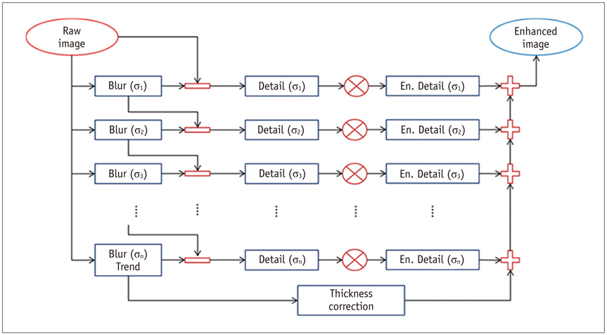
Fig. 2 Principle of multi-scale decompression.
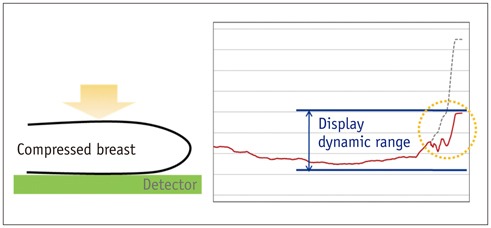
Fig. 3 Effect of thickness correction (global dynamic range reduction).
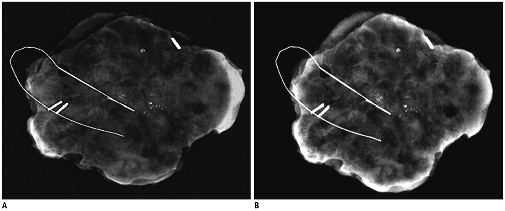
Fig. 4 Specimen mammograms of ductal carcinoma in situ. Mammography (B) was superior to mammography (A) for visualizing calcifications. However, number of calcifications was determined equally well using both mammography protocols. A. Image scanned by specimen mammography without application of "Mammogram enhancement ver. 2.0". B. Image scanned by specimen mammography with application of "Mammogram enhancement ver. 2.0".
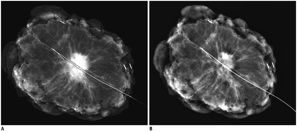
Fig. 5 Specimen mammograms of invasive breast cancer. Mass was visualized similarly using either mammography protocol. A. Image scanned by specimen mammography without application of "Mammogram enhancement ver. 2.0". B. Image scanned by specimen mammography with application of "Mammogram enhancement ver. 2.0".
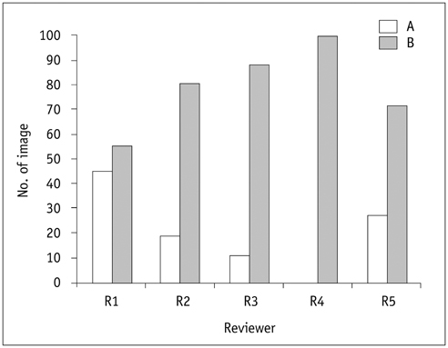
Fig. 6 Graph of priority ranking for five reviewers. A = specimen mammography A, B = specimen mammography B
Cited by 1 articles
-
Performance of Screening Mammography: A Report of the Alliance for Breast Cancer Screening in Korea
Eun Hye Lee, Keum Won Kim, Young Joong Kim, Dong-Rock Shin, Young Mi Park, Hyo Soon Lim, Jeong Seon Park, Hye-Won Kim, You Me Kim, Hye Jung Kim, Jae Kwan Jun
Korean J Radiol. 2016;17(4):489-496. doi: 10.3348/kjr.2016.17.4.489.
Reference
-
1. Meisner AL, Fekrazad MH, Royce ME. Breast disease: benign and malignant. Med Clin North Am. 2008; 92:1115–1141.2. Humphrey LL, Helfand M, Chan BK, Woolf SH. Preventive Services Task Force. Breast cancer screening: a summary of the evidence for the U.S. Ann Intern Med. 2002; 137(5 Part 1):347–360.3. ACR. Cancer Facts & Figures 2012. American Cancer Society;Published 2012. Accessed May 2, 2013. http://www.cancer.org/.4. Pisano ED, Gatsonis C, Hendrick E, Yaffe M, Baum JK, Acharyya S, et al. Diagnostic performance of digital versus film mammography for breast-cancer screening. N Engl J Med. 2005; 353:1773–1783.5. Pisano ED, Hendrick RE, Yaffe MJ, Baum JK, Acharyya S, Cormack JB, et al. Diagnostic accuracy of digital versus film mammography: exploratory analysis of selected population subgroups in DMIST. Radiology. 2008; 246:376–383.6. Bloomquist AK, Yaffe MJ, Pisano ED, Hendrick RE, Mawdsley GE, Bright S, et al. Quality control for digital mammography in the ACRIN DMIST trial: part I. Med Phys. 2006; 33:719–736.7. Sivaramakrishna R, Obuchowski NA, Chilcote WA, Cardenosa G, Powell KA. Comparing the performance of mammographic enhancement algorithms: a preference study. AJR Am J Roentgenol. 2000; 175:45–51.8. Goldstraw EJ, Castellano I, Ashley S, Allen S. The effect of Premium View post-processing software on digital mammographic reporting. Br J Radiol. 2010; 83:122–128.9. Chen B, Wang W, Huang J, Zhao M, Cui G, Xu J, et al. Comparison of tissue equalization, and premium view post-processing methods in full field digital mammography. Eur J Radiol. 2010; 76:73–80.10. del Carmen MG, Halpern EF, Kopans DB, Moy B, Moore RH, Goss PE, et al. Mammographic breast density and race. AJR Am J Roentgenol. 2007; 188:1147–1150.11. Jackson VP, Hendrick RE, Feig SA, Kopans DB. Imaging of the radiographically dense breast. Radiology. 1993; 188:297–301.12. Ko ES, Han BK, Kim SM, Ko EY, Jang M, Lyou CY, et al. Comparison of new and established full-field digital mammography systems in diagnostic performance. Korean J Radiol. 2013; 14:164–170.13. ACR. Mammography Quality Control Manual. Reston, VA: American College of Radiology;1999.14. Laine AF, Schuler S, Fan J, Huda W. Mammographic feature enhancement by multiscale analysis. IEEE Trans Med Imaging. 1994; 13:725–740.15. Strickland RN, Hahn HI. Wavelet transforms for detecting microcalcifications in mammograms. IEEE Trans Med Imaging. 1996; 15:218–229.16. Kallergi M, Clarke LP, Qian W, Gavrielides M, Venugopal P, Berman CG, et al. Interpretation of calcifications in screen/film, digitized, and wavelet-enhanced monitor-displayed mammograms: a receiver operating characteristic study. Acad Radiol. 1996; 3:285–293.17. Li Y, Poulos A, McLean D, Rickard M. A review of methods of clinical image quality evaluation in mammography. Eur J Radiol. 2010; 74:e122–e131.18. Pisano ED, Cole EB, Major S, Zong S, Hemminger BM, Muller KE, et al. Radiologists' preferences for digital mammographic display. The International Digital Mammography Development Group. Radiology. 2000; 216:820–830.19. Schueller G, Riedl CC, Mallek R, Eibenberger K, Langenberger H, Kaindl E, et al. Image quality, lesion detection, and diagnostic efficacy in digital mammography: full-field digital mammography versus computed radiography-based mammography using digital storage phosphor plates. Eur J Radiol. 2008; 67:487–496.20. Skaane P, Balleyguier C, Diekmann F, Diekmann S, Piguet JC, Young K, et al. Breast lesion detection and classification: comparison of screen-film mammography and full-field digital mammography with soft-copy reading--observer performance study. Radiology. 2005; 237:37–44.21. Good WF, Sumkin JH, Dash N, Johns CM, Zuley ML, Rockette HE, et al. Observer sensitivity to small differences: a multipoint rank-order experiment. AJR Am J Roentgenol. 1999; 173:275–278.
- Full Text Links
-
- Actions
-
Cited
- CITED
-
- Close
- Share
-
- Similar articles
-
- Simple post-processing of medical images
- Abdominal Digital Radiography with a Novel Post-Processing Technique: Phantom and Patient Studies
- Nuclear Medical Physics – A Review of an Advanced Technology
- Image enhancement of digital periapical radiographs according to diagnostic tasks
- Comparison Study of Image Quality of Direct and Indirect Conversion Digital Mammography System
