Retrospective study on change in pharyngeal airway space and hyoid bone position after mandibular setback surgery
- Affiliations
-
- 1Division of Oral and Maxillofacial Surgery, Department of Dentistry, Ajou University School of Medicine, Suwon, Korea. seungilsong@hanmail.net
- 2Department of Dentistry, Gumdan Top General Hospital, Incheon, Korea.
- KMID: 2070435
- DOI: http://doi.org/10.5125/jkaoms.2015.41.5.224
Abstract
OBJECTIVES
The purpose of this study was to evaluate changes in the pharyngeal airway space and hyoid bone position after mandibular setback surgery with bilateral sagittal split ramus osteotomy (BSSRO) and to analyze the correlation between the amount of mandibular setback and the amount of change in pharyngeal airway space or hyoid bone position.
MATERIALS AND METHODS
From January 2010 to February 2013, a total of 30 patients who were diagnosed with skeletal class III malocclusion and underwent the same surgery (BSSRO) and fixation method in the Division of Oral and Maxillofacial Surgery, Department of Dentistry at the Ajou University School of Medicine (Suwon, Korea) were included in this study. Lateral cephalograms of the 30 patients were assessed preoperatively (T1), immediately postoperatively (T2), and 6 months postoperatively (T3) to investigate the significance of changes by time and the correlation between the amount of mandibular setback and the amount of change in the airway space and hyoid bone position.
RESULTS
Three regions of the nasopharynx, oropharynx, and hypopharynx were measured and only the oropharynx showed a statistically significant decrease (P<0.01). A significant posterior and inferior displacement of the hyoid bone was found 6 months after surgery (P<0.01). Analysis of the correlation between the amount of mandibular setback and the amount of final change in the airway space and hyoid bone position with Pearson's correlation showed no significant correlation.
CONCLUSION
In this study, the oropharynx significantly decreased after mandibular setback surgery, and changes in the surrounding structures were identified through posteroinferior movement of the hyoid bone during long-term follow-up. Therefore, postoperative obstructive sleep apnea should be considered in patients who plan to undergo mandibular setback surgery, and necessary modifications to the treatment plan should also be considered.
Keyword
MeSH Terms
Figure
-
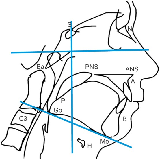
Fig. 1 Parameters and landmarks used for cephalometry. Upper horizontal line: Frankfort horizontal plane, lower oblique line: mandibular plane, vertical line: Sella vertical plane. (S: Sella, N: nasion, Ba: basion, PNS: posterior nasal spine, ANS: anterior nasal spine, A: A point, P: the most inferior point of soft palate, Go: gonion, C3: third cervical vertebra, B: B point, Me: menton, H: hyoid bone)
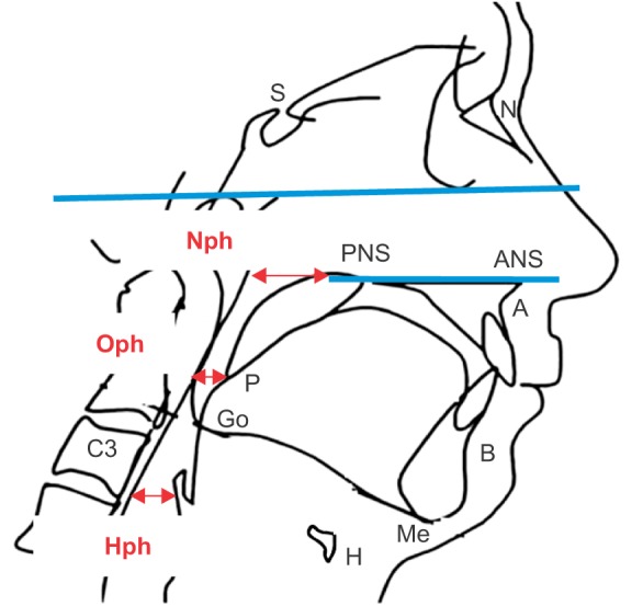
Fig. 2 Measurement of the pharyngeal airway space. The airway space measurement was classified into three areas: the nasopharynx (Nph), oropharynx (Oph), and hypopharynx (Hph). (S: Sella, N: nasion, PNS: posterior nasal spine, ANS: anterior nasal spine, A: A point, P: the most inferior point of soft palate, Go: gonion, C3: third cervical vertebra, B: B point, Me: menton, H: hyoid bone)
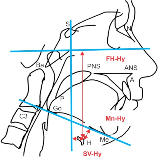
Fig. 3 Measurement of hyoid bone position. The hyoid bone position was evaluated with 3 measuring items using the horizontal position (SV-Hy), vertical position (FH-Hy), and distance to the mandibular inferior border (Mn-Hy). (S: Sella, N: nasion, Ba: basion, PNS: posterior nasal spine, ANS: anterior nasal spine, A: A point, P: the most inferior point of soft palate, Go: gonion, C3: third cervical vertebra, Me: menton, H: hyoid bone)
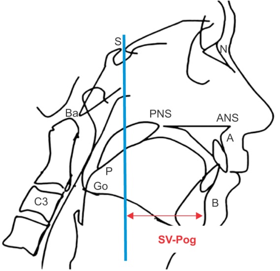
Fig. 4 Measurement of SV-Pog. The vertical distance from the Sella vertical (SV) plane to the pogonion (Pog) was measured to evaluate recurrence. (S: Sella, N: nasion, Ba: basion, PNS: posterior nasal spine, ANS: anterior nasal spine, A: A point, P: the most inferior point of soft palate, Go: gonion, C3: third cervical vertebra, B: B point)
Reference
-
1. Athanasiou AE, Toutountzakis N, Mavreas D, Ritzau M, Wenzel A. Alterations of hyoid bone position and pharyngeal depth and their relationship after surgical correction of mandibular prognathism. Am J Orthod Dentofacial Orthop. 1991; 100:259–265. PMID: 1877552.
Article2. Lee DK, Kim SK. Study on the changes in the upper airway following osteotomy for the mandibular prognathism. J Korean Dent Assoc. 1989; 27:1143–1153.3. Chin KS, Shon WS. The relationships between the postoperative stability and the changes in the tongue position, the hyoid bone position and the upper airway size after orthognathic surgery in patients with mandibular prognathism. Korean J Orthod. 1993; 23:693–705.4. Choi JY, Lee SC. A study on changes of hyoid bone position and upper airway following orthognathic surgery. Kyung Hee Dent J. 1993; 15:729–742.5. Tselnik M, Pogrel MA. Assessment of the pharyngeal airway space after mandibular setback surgery. J Oral Maxillofac Surg. 2000; 58:282–285. PMID: 10716109.
Article6. Greco JM, Frohberg U, Van Sickels JE. Long-term airway space changes after mandibular setback using bilateral sagittal split osteotomy. Int J Oral Maxillofac Surg. 1990; 19:103–105. PMID: 2111357.
Article7. Enacar A, Aksoy AU, Sençift Y, Haydar B, Aras K. Changes in hypopharyngeal airway space and in tongue and hyoid bone positions following the surgical correction of mandibular prognathism. Int J Adult Orthodon Orthognath Surg. 1994; 9:285–290. PMID: 7751760.8. Güven O, Saraçoğlu U. Changes in pharyngeal airway space and hyoid bone positions after body ostectomies and sagittal split ramus osteotomies. J Craniofac Surg. 2005; 16:23–30. PMID: 15699641.
Article9. Park BW, Kim JR. A cephalometric study on changes in pharyngeal airway space, tongue and hyoid bone positions following the surgical correction of mandibular prognathism. J Korean Assoc Oral Maxillofac Surg. 2000; 26:164–171.10. Kawakami M, Yamamoto K, Fujimoto M, Ohgi K, Inoue M, Kirita T. Changes in tongue and hyoid positions, and posterior airway space following mandibular setback surgery. J Craniomaxillofac Surg. 2005; 33:107–110. PMID: 15804589.
Article11. Lew KK. Changes in tongue and hyoid bone positions following anterior mandibular subapical osteotomy in patients with Class III malocclusion. Int J Adult Orthodon Orthognath Surg. 1993; 8:123–128. PMID: 8228428.12. Eggensperger N, Smolka W, Iizuka T. Long-term changes of hyoid bone position and pharyngeal airway size following mandibular setback by sagittal split ramus osteotomy. J Craniomaxillofac Surg. 2005; 33:111–117. PMID: 15804590.
Article13. Choi YH, Kim BK, Choi BJ, Kim YG, Lee BS, Kwon YD, et al. An investigation of hyoid bone position and airway space in class III malocclusion after orthognathic surgery. J Korean Assoc Maxillofac Plast Reconstr Surg. 2011; 33:401–406.14. Riley RW, Powell NB. Maxillofacial surgery and obstructive sleep apnea syndrome. Otolaryngol Clin North Am. 1990; 23:809–826. PMID: 2381716.15. Harris RS. The effect of extension of the head and neck upon the infrahyoid respiratory passage and the supraclavicular portion of the human trachea. Thorax. 1959; 14:176–180. PMID: 14400005.
Article16. Hellsing E. Changes in the pharyngeal airway in relation to extension of the head. Eur J Orthod. 1989; 11:359–365. PMID: 2591483.
Article17. Ono T, Otsuka R, Kuroda T, Honda E, Sasaki T. Effects of head and body position on two- and three-dimensional configurations of the upper airway. J Dent Res. 2000; 79:1879–1884. PMID: 11145359.
Article18. Solow B, Tallgren A. Head posture and craniofacial morphology. Am J Phys Anthropol. 1976; 44:417–435. PMID: 937521.
Article19. Muto T, Takeda S, Kanazawa M, Yamazaki A, Fujiwara Y, Mizoguchi I. The effect of head posture on the pharyngeal airway space (PAS). Int J Oral Maxillofac Surg. 2002; 31:579–583. PMID: 12521311.
Article20. Malkoc S, Usumez S, Nur M, Donaghy CE. Reproducibility of airway dimensions and tongue and hyoid positions on lateral cephalograms. Am J Orthod Dentofacial Orthop. 2005; 128:513–516. PMID: 16214635.
Article21. Partinen M, Guilleminault C, Quera-Salva MA, Jamieson A. Obstructive sleep apnea and cephalometric roentgenograms. The role of anatomic upper airway abnormalities in the definition of abnormal breathing during sleep. Chest. 1988; 93:1199–1205. PMID: 3371099.22. Riley R, Guilleminault C, Herran J, Powell N. Cephalometric analyses and flow-volume loops in obstructive sleep apnea patients. Sleep. 1983; 6:303–311. PMID: 6665392.
Article23. Hochban W, Schürmann R, Brandenburg U, Conradt R. Mandibular setback for surgical correction of mandibular hyperplasia: does it provoke sleep-related breathing disorders? Int J Oral Maxillofac Surg. 1996; 25:333–338. PMID: 8961010.24. Chung DH, Lee KS. A study on changes of airway, tongue, and hyoid position following orthognathic surgery. Korean J Orthod. 1998; 28:487–498.
- Full Text Links
-
- Actions
-
Cited
- CITED
-
- Close
- Share
-
- Similar articles
-
- A Cephalometric Study on Changes in Pharyngeal Airway Space, Tongue and Hyoid Bone Positions Following the Surgical Correction of Mandibular Prognathism
- Changes in the hyoid bone, tongue, and oropharyngeal airway space after mandibular setback surgery evaluated by cone-beam computed tomography
- A cephalometric study on changes in hyoid bone, tongue and upper airway space according to skeletal change in persons with mandible prognathism after orthognathic surgery
- Retrospective study of changes in pharyngeal airway space and position of hyoid bone after mandibular setback surgery by cephalometric analysis
- The effects of mandibular setback osteotomy on the oropharyngeal airway space in mandibular prognathic patients
