Feasibility of utilizing the patellar ligament angle for assessing cranial cruciate ligament rupture in dogs
- Affiliations
-
- 1Department of Veterinary Surgery, College of Veterinary Medicine, Konkuk University, Seoul 143-701, Korea. swjeong@konkuk.ac.kr
- KMID: 2070244
- DOI: http://doi.org/10.4142/jvs.2014.15.4.563
Abstract
- The patellar ligament angle (PLA) was assessed in 105 normal stifle joints of 79 dogs and 33 stifle joints of 26 dogs with a ruptured cranial cruciate ligament (CrCL). The PLA of stifles with complete CrCL rupture was significantly lower than that of normal stifles, particularly at a flexion angle of 60~80degrees in both plain and stress views. If the PLA was <90.55degrees on the stress view with a 60~80degrees flexion angle, the dog was diagnosed with a complete rupture of the CrCL with a sensitivity of 83.9% and specificity of 100%. In conclusion, measuring the PLA is a quantitative method for diagnosing complete CrCL rupture in canines.
MeSH Terms
Figure
-
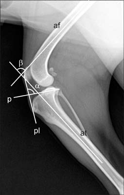
Fig. 1 Mediolateral radiographic view of a dog with a normal stifle joint to illustrate measuring the angles α and β. Angle α was defined as the angle between the patellar ligament (pl) and tibial plateau (p). The flexion angle (β) of the stifle joint was measured between the long axis of the femur (af) and long axis of the tibia (at).
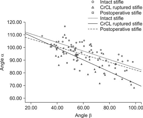
Fig. 2 Inclination of angle α at the incident stifle flexion along with angle β in dogs with complete rupture of the CrCL (triangles), after surgery (squares), and normal stifles (circles). The equation of the linear regression analysis for α vs. β before surgery was as follows: α = -0.78β + 118 (r2 = 0.602; p < 0.001; continuous line). The equation of the linear regression analysis for α vs. β after surgery was as follows: α = -0.56β + 113 (r2 = 0.314; p < 0.001; wide dashed line). The small dashed line is the regression line for normal dogs: α = -0.32β + 116 (r2 = 0.416; p < 0.001).
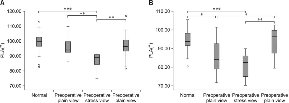
Fig. 3 Comparison of the patellar ligament angle (PLA) at a 40~60° (A) or 60~80° (B) flexion angle. Differences were considered to be significant at *p < 0.05, **p < 0.01, and ***p < 0.001.
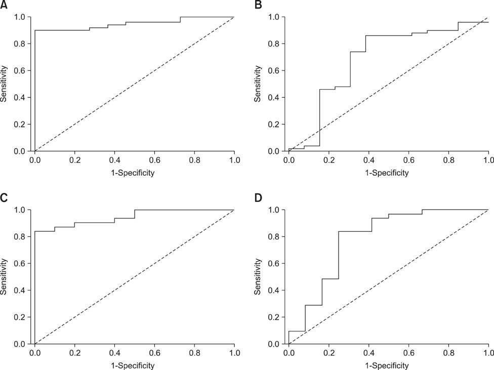
Fig. 4 Receiver operating characteristic (ROC) curves for the PLA with a 40~60° flexion angle on the stress (A) and plain views (B), and with a 60~80° flexion angle on the stress (C) and plain views (D). (A) The area under the curve (AUC) was 0.949. With a 92.40° cutoff value, the PLA measurement had a sensitivity of 90.0% and specificity of 100%. (B) The AUC was 0.686. With a 94.51° cutoff value, the PLA measurement had a sensitivity of 86.0% and specificity of 61.5%. (C) The AUC was 0.945. With a 90.55° cutoff value, the PLA measurement had a sensitivity of 83.9% and specificity of 100%. (D) The AUC was 0.785. With a 90.76° cutoff value, the PLA measurement had a sensitivity of 83.9% and specificity of 75.0%.
Reference
-
1. Butler DL, Noyes FR, Grood ES. Ligamentous restraints to anterior-posterior drawer in the human knee. A biomechenical study. J Bone Joint Surg Am. 1980; 62:259–270.
Article2. Dennler R, Kipfer NM, Tepic S, Hassig M, Montavon PM. Inclination of the patellar ligament in relation to flexion angle in stifle joints of dogs without degenerative joint disease. Am J Vet Res. 2006; 67:1849–1854.
Article3. Havig ME, Dyce J, Kowaleski MP, Reynolds LR, Budsberg SC. Relationship of tibial plateau slope to limb function in dogs treated with a lateral suture technique for stabilization of cranial cruciate ligament deficient stifles. Vet Surg. 2007; 36:245–251.
Article4. Might KR, Bachelez A, Martinez SA, Gay JM. Evaluation of the drawer test and the tibial compression test for differentiating between cranial and caudal stifle subluxation associated with cruciate ligament instability. Vet Surg. 2013; 42:392–397.
Article5. Nisell R. Mechanics of the knee. A study of joint and muscle load with clinical applications. Acta Orthop Scand Suppl. 1985; 216:1–42.6. Pond MJ, Campbell JR. The canine stifle joint I. Rupture of the anterior cruciate ligament. An assessment of conservative and surgical treatment. J Small Anim Pract. 1972; 13:1–10.7. de Rooster H, Van Ryssen B, van Bree H. Diagnosis of cranial cruciate ligament injury in dogs by tibial compression radiography. Vet Rec. 1998; 142:366–368.
Article8. Schwandt CS, Bohorquez-Vanelli A, Tepic S, Hassig M, Dennler R, Vezzoni A, Montavon PM. Angle between the patellar ligament and tibial plateau in dogs with partial rupture of the cranial cruciate ligament. Am J Vet Res. 2006; 67:1855–1860.
Article
- Full Text Links
-
- Actions
-
Cited
- CITED
-
- Close
- Share
-
- Similar articles
-
- Patellar Tendon Rupture associated with Rupture of Anterior Cruciate Ligament, Both Collateral Ligoments, and Lateral Meniscus: A Case Report
- Acute Simultaneous Ruptures of the Anterior Cruciate Ligament and Patellar Tendon
- Evaluation of partial cranial cruciate ligament rupture with positive contrast computed tomographic arthrography in dogs
- Reconstruction of Posterior Cruciate Ligament Using Semitendinosus Tendon (3 cases)
- Anterior Cruciate Ligament Reconstruction using Patellar tendon with Kennedy-LAD
