Qualitative and Quantitative Analysis with Contrast-Enhanced Ultrasonography: Diagnosis Value in Hypoechoic Renal Angiomyolipoma
- Affiliations
-
- 1Shanghai Imaging Institute of Medicine, Department of Ultrasound, Zhongshan Hospital, Fudan University, Shanghai 200032, China. huang.beijian@zs-hospital.sh.cn
- KMID: 2070177
- DOI: http://doi.org/10.3348/kjr.2015.16.2.334
Abstract
OBJECTIVE
To evaluate the value of enhancement features and quantitative parameters of contrast-enhanced ultrasonography (CEUS) in differentiating solid hypoechoic renal angiomyolipomas (AMLs) from clear cell renal cell carcinomas (ccRCCs).
MATERIALS AND METHODS
We analyzed the enhancement features and quantitative parameters of CEUS in 174 hypoechoic renal masses (32 AMLs and 142 ccRCCs) included in the study.
RESULTS
Centripetal enhancement pattern was more common in AMLs than in ccRCCs on CEUS (71.9% vs. 23.2%, p < 0.001). At peak enhancement, all AMLs showed homogeneous enhancement (100% in AML, 27.5% in ccRCCs; p < 0.001). Quantitative analysis showed no significant difference between rise time and time to peak. Tumor-to-cortex (TOC) enhancement ratio in AMLs was significantly lower than that in ccRCCs (p < 0.001). The criteria of centripetal enhancement and homogeneous peak enhancement together with TOC ratio < 91.0% used to differentiate hypoechoic AMLs from ccRCCs resulted in a sensitivity and specificity of 68.9% and 95.8%, respectively.
CONCLUSION
Both qualitative and quantitative analysis with CEUS are valuable in the differential diagnosis of hypoechoic renal AMLs from ccRCCs.
Keyword
MeSH Terms
-
Adult
Aged
Angiomyolipoma/*diagnosis/pathology/*ultrasonography
Carcinoma, Renal Cell/*diagnosis/pathology/*ultrasonography
Contrast Media
Diagnosis, Differential
Female
Humans
Kidney Neoplasms/diagnosis/pathology/*ultrasonography
Language
Lipoma/ultrasonography
Male
Middle Aged
Sensitivity and Specificity
Contrast Media
Figure
-
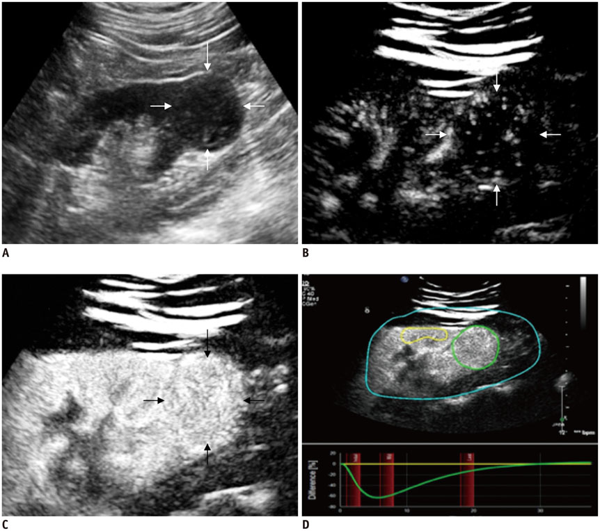
Fig. 1 Epithelioid angiomyolipoma (arrows) in 28-year-old man. A. Ultrasonography shows 35 × 34 mm solid hypoechoic lesion in low pole of right kidney. B. At 14 seconds after injection of Sonovue, lesion shows centripetal enhancement. C. At 22 seconds, lesion shows homogeneous peak enhancement. D. Quantitative analysis with CEUS shows TOC ratio of 61.7%. Enhancement degree of lesion is lower than that of adjacent renal cortex (yellow, reference ROI; green, analysis ROI). CEUS = contrast-enhanced ultrasonography, ROI = regions of interest, TOC ratio = tumor-to-cortex enhancement ratio
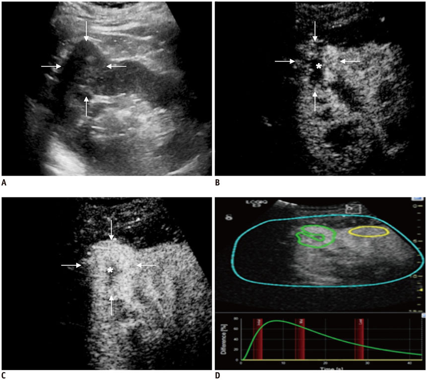
Fig. 2 Clear cell renal cell carcinoma (arrows) in 65-year-old man. A. Ultrasonography shows 35 × 32 mm solid hypoechoic lesion, located in upper pole of right kidney. B. At 16 seconds time point post-injection of Sonovue, lesion shows entire enhancement with inner non-enhanced area (asterisk). C. Lesion shows peak enhancement at 22 seconds, with non-enhanced area throughout CEUS progress (asterisk). D. Quantitative analysis with CEUS shows TOC ratio of 178.1%. Enhancement degree of lesion is higher than that of adjacent renal cortex (yellow, reference ROI; green, analysis ROI, encompassing enhanced area as much as possible with irregular shape). CEUS = contrast-enhanced ultrasonography, ROI = regions of interest, TOC ratio = tumor-to-cortex enhancement ratio

Fig. 3 CEUS shows incomplete pseudocapsule sign (arrows) around homogeneous enhanced lesion. A. 36 × 34 mm epithelioid minimal-fat renal angiomyolipoma in upper pole of right kidney in 43-year-old woman. At 18 seconds time point post-injection of Sonovue. B. 41 × 40 mm epithelioid minimal-fat renal angiomyolipoma in middle pole of left kidney in 52-year-old man. At 20 seconds time point post-injection of Sonovue. CEUS = contrast-enhanced ultrasonography
Cited by 1 articles
-
Dynamic Contrast-Enhanced Ultrasound of Gastric Cancer: Correlation with Perfusion CT and Histopathology
Ijin Joo, Se Hyung Kim, Dong Ho Lee, Joon Koo Han
Korean J Radiol. 2019;20(5):781-790. doi: 10.3348/kjr.2018.0273.
Reference
-
1. Hartman DS, Goldman SM, Friedman AC, Davis CJ Jr, Madewell JE, Sherman JL. Angiomyolipoma: ultrasonic-pathologic correlation. Radiology. 1981; 139:451–458.2. Halpenny D, Snow A, McNeill G, Torreggiani WC. The radiological diagnosis and treatment of renal angiomyolipoma-current status. Clin Radiol. 2010; 65:99–108.3. Lu Q, Wang W, Huang B, Li C, Li C. Minimal fat renal angiomyolipoma: the initial study with contrast-enhanced ultrasonography. Ultrasound Med Biol. 2012; 38:1896–1901.4. Aydin H, Magi-Galluzzi C, Lane BR, Sercia L, Lopez JI, Rini BI, et al. Renal angiomyolipoma: clinicopathologic study of 194 cases with emphasis on the epithelioid histology and tuberous sclerosis association. Am J Surg Pathol. 2009; 33:289–297.5. Serrano Frago P, Del Agua Arias Camisón C, Gil Sanz MJ, Allué López M, Gonzalvo Ibarra A, Plaza Mas L, et al. Controversies related to epithelioid variant of renal angiomyolipoma: a review of the literature. Urology. 2006; 67:846.e3. 846.e5.6. Xu ZF, Xu HX, Xie XY, Liu GJ, Zheng YL, Lu MD. Renal cell carcinoma and renal angiomyolipoma: differential diagnosis with real-time contrast-enhanced ultrasonography. J Ultrasound Med. 2010; 29:709–717.7. Ascenti G, Gaeta M, Magno C, Mazziotti S, Blandino A, Melloni D, et al. Contrast-enhanced second-harmonic sonography in the detection of pseudocapsule in renal cell carcinoma. AJR Am J Roentgenol. 2004; 182:1525–1530.8. Pretorius ES, Siegelman ES, Ramchandani P, Cangiano T, Banner MP. Renal neoplasms amenable to partial nephrectomy: MR imaging. Radiology. 1999; 212:28–34.9. Ignee A, Jedrejczyk M, Schuessler G, Jakubowski W, Dietrich CF. Quantitative contrast enhanced ultrasound of the liver for time intensity curves-reliability and potential sources of errors. Eur J Radiol. 2010; 73:153–158.10. Raj GV, Bach AM, Iasonos A, Korets R, Blitstein J, Hann L, et al. Predicting the histology of renal masses using preoperative Doppler ultrasonography. J Urol. 2007; 177:53–58.11. Milner J, McNeil B, Alioto J, Proud K, Rubinas T, Picken M, et al. Fat poor renal angiomyolipoma: patient, computerized tomography and histological findings. J Urol. 2006; 176:905–909.12. Sasiwimonphan K, Takahashi N, Leibovich BC, Carter RE, Atwell TD, Kawashima A. Small (< 4 cm) renal mass: differentiation of angiomyolipoma without visible fat from renal cell carcinoma utilizing MR imaging. Radiology. 2012; 263:160–168.13. Pysz MA, Guracar I, Foygel K, Tian L, Willmann JK. Quantitative assessment of tumor angiogenesis using real-time motion-compensated contrast-enhanced ultrasound imaging. Angiogenesis. 2012; 15:433–442.14. Goertz RS, Bernatik T, Strobel D, Hahn EG, Haendl T. Software-based quantification of contrast-enhanced ultrasound in focal liver lesions--a feasibility study. Eur J Radiol. 2010; 75:e22–e26.15. Aoki S, Hattori R, Yamamoto T, Funahashi Y, Matsukawa Y, Gotoh M, et al. Contrast-enhanced ultrasound using a time-intensity curve for the diagnosis of renal cell carcinoma. BJU Int. 2011; 108:349–354.16. Tsai CC, Wu WJ, Li CC, Wang CJ, Wu CH, Wu CC. Epithelioid angiomyolipoma of the kidney mimicking renal cell carcinoma: a clinicopathologic analysis of cases and literature review. Kaohsiung J Med Sci. 2009; 25:133–140.17. Park BK, Kim SH, Choi HJ. Characterization of renal cell carcinoma using agent detection imaging: comparison with gray-scale US. Korean J Radiol. 2005; 6:173–178.18. Jiang J, Chen Y, Zhou Y, Zhang H. Clear cell renal cell carcinoma: contrast-enhanced ultrasound features relation to tumor size. Eur J Radiol. 2010; 73:162–167.19. Dong XQ, Shen Y, Xu LW, Xu CM, Bi W, Wang XM. Contrast-enhanced ultrasound for detection and diagnosis of renal clear cell carcinoma. Chin Med J (Engl). 2009; 122:1179–1183.20. Gerst S, Hann LE, Li D, Gonen M, Tickoo S, Sohn MJ, et al. Evaluation of renal masses with contrast-enhanced ultrasound: initial experience. AJR Am J Roentgenol. 2011; 197:897–906.21. Yamashita Y, Honda S, Nishiharu T, Urata J, Takahashi M. Detection of pseudocapsule of renal cell carcinoma with MR imaging and CT. AJR Am J Roentgenol. 1996; 166:1151–1155.22. Ruppert-Kohlmayr AJ, Uggowitzer M, Meissnitzer T, Ruppert G. Differentiation of renal clear cell carcinoma and renal papillary carcinoma using quantitative CT enhancement parameters. AJR Am J Roentgenol. 2004; 183:1387–1391.23. Sheir KZ, El-Azab M, Mosbah A, El-Baz M, Shaaban AA. Differentiation of renal cell carcinoma subtypes by multislice computerized tomography. J Urol. 2005; 174:451–455. discussion 455.24. Siracusano S, Quaia E, Bertolotto M, Ciciliato S, Tiberio A, Belgrano E. The application of ultrasound contrast agents in the characterization of renal tumors. World J Urol. 2004; 22:316–322.25. Zhang J, Lefkowitz RA, Ishill NM, Wang L, Moskowitz CS, Russo P, et al. Solid renal cortical tumors: differentiation with CT. Radiology. 2007; 244:494–504.26. Pedrosa I, Chou MT, Ngo L, H Baroni R, Genega EM, Galaburda L, et al. MR classification of renal masses with pathologic correlation. Eur Radiol. 2008; 18:365–375.27. Kim JK, Kim TK, Ahn HJ, Kim CS, Kim KR, Cho KS. Differentiation of subtypes of renal cell carcinoma on helical CT scans. AJR Am J Roentgenol. 2002; 178:1499–1506.
- Full Text Links
-
- Actions
-
Cited
- CITED
-
- Close
- Share
-
- Similar articles
-
- A Case of Renal Angiomyolipoma
- Renal Angiomyolipoma with the Regional Lymph Nodes Involvement
- Hepatic Hemangioma with Hypoechoic Halo Accompanied by Arterioportal Shunt
- A Case of Renal Angiomyolipoma
- Circularity Index on Contrast-Enhanced Computed Tomography Helps Distinguish Fat-Poor Angiomyolipoma from Renal Cell Carcinoma: Retrospective Analyses of Histologically Proven 257 Small Renal Tumors Less Than 4 cm
