How to Minimize Rotational Conflict between Femoral & Tibial Component in Total Knee Arthroplasty: The Use of Femoro-Tibial Axial Synchronizer (Linker)
- Affiliations
-
- 1Department of Orthopaedic Surgery, Samsung Medical Center, Sungkyunkwan University School of Medicine, Seoul, Korea.
- 2Department of Orthopaedic Surgery, National Health Insurance Service Ilsan Hospital, Goyang, Korea. orthomania@gmail.com
- KMID: 2070024
- DOI: http://doi.org/10.3349/ymj.2015.56.2.454
Abstract
- PURPOSE
The purpose of this study was to investigate the correlation between rotational axes of femur and tibia with the use of Linker.
MATERIALS AND METHODS
This study was carried out from August 2009 to February 2010 on 54 patients (106 knees), who were diagnosed with simultaneous bilateral total knee arthroplasty. With the use of postoperative computed tomography scans, it was investigated how much the rotational angle of femoral and tibial components matched.
RESULTS
The tibial component was internally rotated for the femoral component at an angle of 0.8degrees. The femoral component was externally rotated for the surgical transepicondylar axis (TEA) at an angle of 1.6 (range: from 4.8degrees of internal rotation to 7.9degrees of external rotation, SD=2.2degrees), and the tibial component was externally rotated for the surgical TEA at an average angle of 0.9 (range: from 5.1degrees of internal rotation to 8.3degrees of external rotation, SD=3.1degrees).
CONCLUSION
The femoro-tibial synchronizer helped to improve the orientation and positioning of both femoral component and tibial component, and also increase the correlation of the rotational axes of the two components.
Keyword
MeSH Terms
Figure
-

Fig. 1 Proximal resector with connecting instrument. This tibial AP axis synchronizer is composed of tail, caliper, and proximal tibial resector.

Fig. 2 The photo of distal femoral resector. Linker is composed of distal femoral resector and proximal tibial resector at knee extension position.

Fig. 3 Linker is a connecting instrument of distal femoral resector and proximal tibial resector. When femeur's AP pin is parallel to tibia's AP pin, femoral component has a parallel relationship with a tibial component. And resectional planes have the rectangularity with each other.

Fig. 4 With knee extension position, the synchronization of AP axes of femur and tibia is confirmed. The tail of proximal tibial resector presenting femur's coronal & sagittal axis is synchronized with tibia's coronal & sagittal axis.

Fig. 5 Tibial plate is inserted as synchronized with marked tibial AP axis.
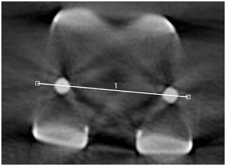
Fig. 6 With the patient supine on the CT scanning table, lower extremities were stabilized in maximum extension in a plastic frame without any internal or external rotation. Femoral component rotational axis was defined as the line joining the posterior margins of both pegs of the femoral component.
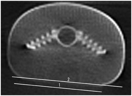
Fig. 7 Rotation of tibial component can be measured with the line connecting the posterior margin of keels.
Cited by 1 articles
-
Revision Arthroplasty Using a MUTARS® Prosthesis in Comminuted Periprosthetic Fracture of the Distal Femur
Hyung-Suk Choi, Jae-Hwi Nho, Chung-Hyun Kim, Sai-Won Kwon, Jong-Seok Park, You-Sung Suh
Yonsei Med J. 2016;57(6):1517-1522. doi: 10.3349/ymj.2016.57.6.1517.
Reference
-
1. Aglietti P, Sensi L, Cuomo P, Ciardullo A. Rotational position of femoral and tibial components in TKA using the femoral transepicondylar axis. Clin Orthop Relat Res. 2008; 466:2751–2755.
Article2. Nicoll D, Rowley DI. Internal rotational error of the tibial component is a major cause of pain after total knee replacement. J Bone Joint Surg Br. 2010; 92:1238–1244.
Article3. Toms AD, Mandalia V, Haigh R, Hopwood B. The management of patients with painful total knee replacement. J Bone Joint Surg Br. 2009; 91:143–150.
Article4. Hirschmann MT, Konala P, Amsler F, Iranpour F, Friederich NF, Cobb JP. The position and orientation of total knee replacement components: a comparison of conventional radiographs, transverse 2D-CT slices and 3D-CT reconstruction. J Bone Joint Surg Br. 2011; 93:629–633.5. Matziolis G, Krocker D, Weiss U, Tohtz S, Perka C. A prospective, randomized study of computer-assisted and conventional total knee arthroplasty. Three-dimensional evaluation of implant alignment and rotation. J Bone Joint Surg Am. 2007; 89:236–243.
Article6. Olcott CW, Scott RD. The Ranawat Award. Femoral component rotation during total knee arthroplasty. Clin Orthop Relat Res. 1999; 39–42.7. Lee DH, Seo JG, Moon YW. Synchronisation of tibial rotational alignment with femoral component in total knee arthroplasty. Int Orthop. 2008; 32:223–227.
Article8. Seo JG, Moon YW, Lim JS, Park SJ, Kim SM. Mechanical axis-derived femoral component rotation in extramedullary total knee arthroplasty: a comparison between femoral transverse axis and transepicondylar axis. Knee Surg Sports Traumatol Arthrosc. 2012; 20:538–545.
Article9. Ishii Y, Terajima K, Koga Y, Bechtold JE. Screw home motion after total knee replacement. Clin Orthop Relat Res. 1999; 181–187.
Article10. Ikeuchi M, Yamanaka N, Okanoue Y, Ueta E, Tani T. Determining the rotational alignment of the tibial component at total knee replacement: a comparison of two techniques. J Bone Joint Surg Br. 2007; 89:45–49.11. Thompson JA, Hast MW, Granger JF, Piazza SJ, Siston RA. Biomechanical effects of total knee arthroplasty component malrotation: a computational simulation. J Orthop Res. 2011; 29:969–975.
Article12. Suter T, Zanetti M, Schmid M, Romero J. Reproducibility of measurement of femoral component rotation after total knee arthroplasty using computer tomography. J Arthroplasty. 2006; 21:744–748.
Article13. Vanin N, Panzica M, Dikos G, Krettek C, Hankemeier S. Rotational alignment in total knee arthroplasty: intraoperative inter- and intraobserver reliability of Whiteside's line. Arch Orthop Trauma Surg. 2011; 131:1477–1480.
Article14. Victor J. Rotational alignment of the distal femur: a literature review. Orthop Traumatol Surg Res. 2009; 95:365–372.
Article15. Won YY, Cui WQ, Baek MH, Yun TB, Han SH. An additional reference axis for determining rotational alignment of the femoral component in total knee arthroplasty. J Arthroplasty. 2007; 22:1049–1053.
Article16. Matziolis G, Pfiel S, Wassilew G, Boenicke H, Perka C. Kinematic analysis of the flexion axis for correct femoral component placement. Knee Surg Sports Traumatol Arthrosc. 2011; 19:1504–1509.
Article17. Merican AM, Ghosh KM, Iranpour F, Deehan DJ, Amis AA. The effect of femoral component rotation on the kinematics of the tibiofemoral and patellofemoral joints after total knee arthroplasty. Knee Surg Sports Traumatol Arthrosc. 2011; 19:1479–1487.
Article18. Harvie P, Sloan K, Beaver RJ. Three-dimensional component alignment and functional outcome in computer-navigated total knee arthroplasty: a prospective, randomized study comparing two navigation systems. J Arthroplasty. 2011; 26:1285–1290.19. Akagi M, Mori S, Nishimura S, Nishimura A, Asano T, Hamanishi C. Variability of extraarticular tibial rotation references for total knee arthroplasty. Clin Orthop Relat Res. 2005; 172–176.
Article20. Akagi M, Oh M, Nonaka T, Tsujimoto H, Asano T, Hamanishi C. An anteroposterior axis of the tibia for total knee arthroplasty. Clin Orthop Relat Res. 2004; 1:213–219.
Article
- Full Text Links
-
- Actions
-
Cited
- CITED
-
- Close
- Share
-
- Similar articles
-
- Accuracy of Limb Alignment in Total Knee Arthroplasty using Image-Free Navigation System: Comparison with Conventional Total Knee Arthroplasty
- Self-aligned Technique for Tibial Component Placement in Total Knee Arthroplasty Lessening Rotational Malalignment in Measured Resection and Gap-Balancing Techniques
- Combined Rotational Alignment Change after Total Knee Arthroplasty in Different Tibial Component Designs: Implications for Optimal Tibial Component Rotational Alignment
- The Accuracy of Lower Extremity Alignment in a Total Knee Arthroplasty Using Computer-Assisted Navigation System
- Total Knee Arthroplasty using Intramedullary Tibial Cutting Guide
