Pedicle Screw Placement in the Thoracolumbar Spine Using a Novel, Simple, Safe, and Effective Guide-Pin : A Computerized Tomography Analysis
- Affiliations
-
- 1Department of Neurosurgery, Spine Center, Seoul National University Bundang Hospital, Seoul National University College of Medicine, Seongnam, Korea.
- 2Department of Orthopaedic Surgery, Spine Service, Columbia University College of Physicians and Surgeons, New York, NY, USA.
- 3Department of Neurosurgery, Asan Medical Center, University of Ulsan College of Medicine, Seoul, Korea. scrhim@amc.seoul.kr
- 4Department of Orthopedic Surgery, Wooridul Spine Hospital, Seoul, Korea.
- 5Department of Orthopaedic Surgery, Icahn School of Medicine at Mount Sinai, New York, NY, USA.
- KMID: 2067098
- DOI: http://doi.org/10.3340/jkns.2015.58.1.9
Abstract
OBJECTIVE
To improve pedicle screw placement accuracy with minimal radiation and low cost, we developed specially designed K-wire with a marker. To evaluate the accuracy of thoracolumbar pedicle screws placed using the novel guide-pin and portable X-rays.
METHODS
Observational cohort study with computerized tomography (CT) analysis of in vivo and in vitro pedicle screw placement. Postoperative CT scans of 183 titanium pedicle screws (85 lumbar and 98 thoracic from T1 to L5) placed into 2 cadavers and 18 patients were assessed. A specially designed guide-pin with a marker was inserted into the pedicle to identify the correct starting point (2 mm lateral to the center of the pedicle) and aiming point (center of the pedicle isthmus) in posteroanterior and lateral X-rays. After radiographically confirming the exact starting and aiming points desired, a gearshift was inserted into the pedicle from the starting point into the vertebral body through the center of pedicle isthmus.
RESULTS
Ninety-nine percent (181/183) of screws were contained within the pedicle (total 183 pedicle screws : 98 thoracic pedicle screws and 85 lumbar screws). Only two of 183 (1.0%) thoracic pedicle screws demonstrated breach (1 lateral in a patient and 1 medial in a cadaver specimen). None of the pedicle breaches were associated with neurologic or other clinical sequelae.
CONCLUSION
A simple, specially designed guide-pin with portable X-rays can provide correct starting and aiming points and allows for accurate pedicle screw placement without preoperative CT scan and intraoperative fluoroscopic assistance.
Keyword
Figure
-
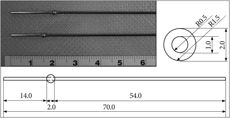
Fig. 1 Photograph (upper left) and schematic illustrations show the precise dimensions of the specially designed guide-pin with a ball maker. The guide-pin is composed of stainless steel. The guide-pin with a ball maker defines the ideal starting point by a ball marker located 15 or 20 mm proximal to the tip and the aiming spot by the tip of the guide-pin.
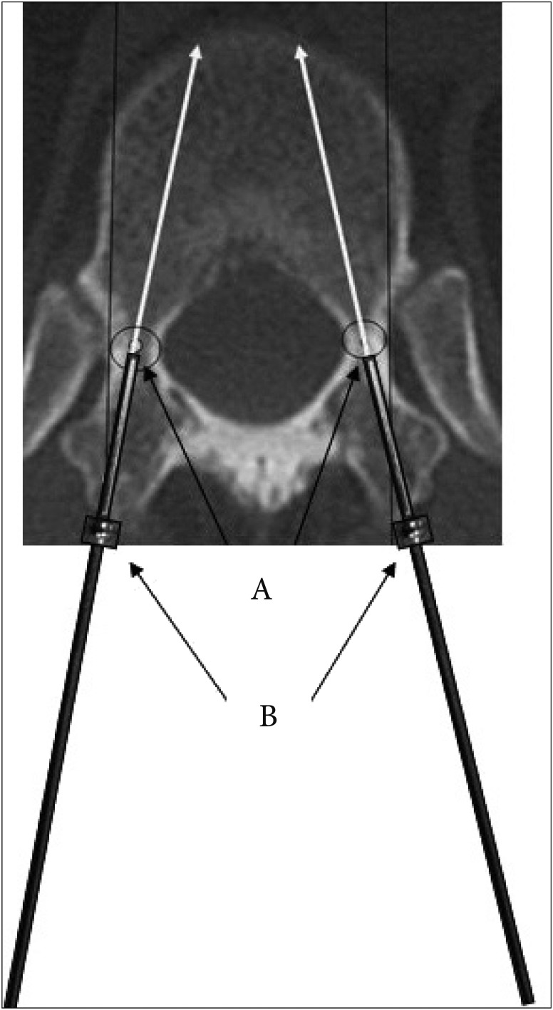
Fig. 2 A novel guide-pin was inserted into the pedicle to identify the correct starting point (A : the laminar 2-3 mm lateral to the center of the pedicle) and aiming spot (B : center of the pedicle isthmus).

Fig. 3 Steps required for pedicle screw fixation with the specially designed guide-pin. Step 1 : complete exposure of the bony anatomy; step 2 : starting point burr (2 mm) with alleged recommendation; step 2-1 : Define the ideal starting point and aiming spot using C-arm or portable postero anterior/lateral X-rays; step 3 : the gearshift insertion according to the X-rays; step 4 : inner pedicle palpation and length measurement; step 5 : tapping and repalpation; and step 6 : screw placement slowly.
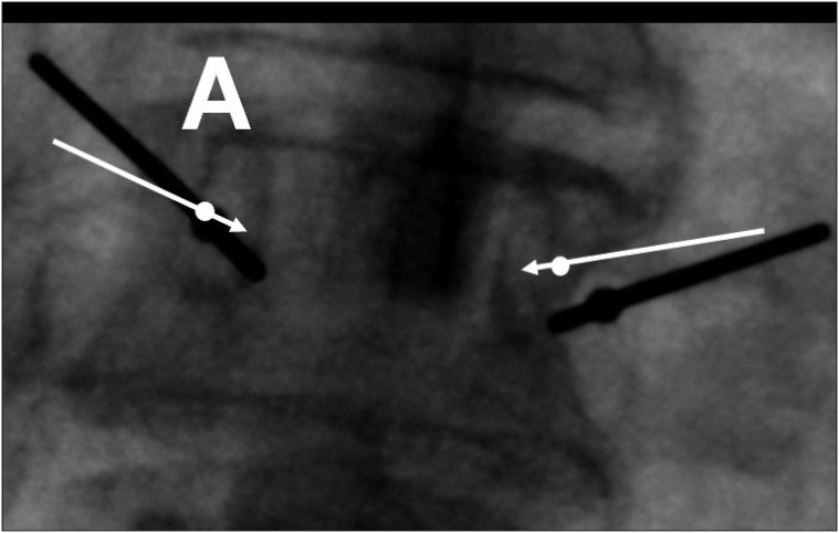
Fig. 4 Adjusting the starting point and aiming spot after complete placement of guide-pin. Guide-pin A : starting point (ball marker) looks okay (2-3 mm lateral to the center of the pedicle) but tip of the guide pin violated medial wall of the pedicle. Ideal targeting (arrows) : same starting point but near perpendicular gearshift insertion required.

Fig. 5 Coronal and sagittal plane radiographs should confirm the harmonious position of the screws by the surgeon.
Reference
-
1. Amiot LP, Lang K, Putzier M, Zippel H, Labelle H. Comparative results between conventional and computer-assisted pedicle screw installation in the thoracic, lumbar, and sacral spine. Spine (Phila Pa 1976). 2000; 25:606–614. PMID: 10749638.
Article2. Belmont PJ Jr, Klemme WR, Dhawan A, Polly DW Jr. In vivo accuracy of thoracic pedicle screws. Spine (Phila Pa 1976). 2001; 26:2340–2346. PMID: 11679819.
Article3. Brown CA, Lenke LG, Bridwell KH, Geideman WM, Hasan SA, Blanke K. Complications of pediatric thoracolumbar and lumbar pedicle screws. Spine (Phila Pa 1976). 1998; 23:1566–1571. PMID: 9682313.
Article4. Cotrel Y, Dubousset J, Guillaumat M. New universal instrumentation in spinal surgery. Clin Orthop Relat Res. 1988; 227:10–23. PMID: 3338200.
Article5. Esses SI, Sachs BL, Dreyzin V. Complications associated with the technique of pedicle screw fixation. A selected survey of ABS members. Spine (Phila Pa 1976). 1993; 18:2231–2238. discussion 2238-2239PMID: 8278838.
Article6. Farber GL, Place HM, Mazur RA, Jones DE, Damiano TR. Accuracy of pedicle screw placement in lumbar fusions by plain radiographs and computed tomography. Spine (Phila Pa 1976). 1995; 20:1494–1499. PMID: 8623069.
Article7. Ferrick MR, Kowalski JM, Simmons ED Jr. Reliability of roentgenogram evaluation of pedicle screw position. Spine (Phila Pa 1976). 1997; 22:1249–1252. discussion 1253PMID: 9201864.
Article8. Hyun SJ, Kim YJ, Cheh G, Yoon SH, Rhim SC. Free hand pedicle screw placement in the thoracic spine without any radiographic guidance : technical note, a cadaveric study. J Korean Neurosurg Soc. 2012; 51:66–70. PMID: 22396849.
Article9. Kim KD, Johnson JP, Babbitz JD. Image-guided thoracic pedicle screw placement : a technical study in cadavers and preliminary clinical experience. Neurosurg Focus. 2001; 10:E2. PMID: 16749749.10. Kim YJ, Lenke LG, Bridwell KH, Cho YS, Riew KD. Free hand pedicle screw placement in the thoracic spine : is it safe? Spine (Phila Pa 1976). 2004; 29:333–342. discussion 342PMID: 14752359.11. Kim YJ, Lenke LG, Cheh G, Riew KD. Evaluation of pedicle screw placement in the deformed spine using intraoperative plain radiographs : a comparison with computerized tomography. Spine (Phila Pa 1976). 2005; 30:2084–2088. PMID: 16166900.
Article12. Kuntz C 4th, Maher PC, Levine NB, Kurokawa R. Prospective evaluation of thoracic pedicle screw placement using fluoroscopic imaging. J Spinal Disord Tech. 2004; 17:206–214. PMID: 15167336.
Article13. Laine T, Mäkitalo K, Schlenzka D, Tallroth K, Poussa M, Alho A. Accuracy of pedicle screw insertion : a prospective CT study in 30 low back patients. Eur Spine J. 1997; 6:402–405. PMID: 9455669.
Article14. Lee CH, Hyun SJ, Kim YJ, Kim KJ, Jahng TA, Kim HJ. Accuracy of free hand pedicle screw installation in the thoracic and lumbar spine by a young surgeon : an analysis of the first consecutive 306 screws using computed tomography. Asian Spine J. 2014; 8:237–243. PMID: 24967036.
Article15. Li G, Lv G, Passias P, Kozanek M, Metkar US, Liu Z, et al. Complications associated with thoracic pedicle screws in spinal deformity. Eur Spine J. 2010; 19:1576–1584. PMID: 20237943.
Article16. Liljenqvist UR, Halm HF, Link TM. Pedicle screw instrumentation of the thoracic spine in idiopathic scoliosis. Spine (Phila Pa 1976). 1997; 22:2239–2245. PMID: 9346144.
Article17. Liljenqvist UR, Link TM, Halm HF. Morphometric analysis of thoracic and lumbar vertebrae in idiopathic scoliosis. Spine (Phila Pa 1976). 2000; 25:1247–1253. PMID: 10806501.
Article18. Lonstein JE, Denis F, Perra JH, Pinto MR, Smith MD, Winter RB. Complications associated with pedicle screws. J Bone Joint Surg Am. 1999; 81:1519–1528. PMID: 10565643.
Article19. Rajasekaran S, Vidyadhara S, Ramesh P, Shetty AP. Randomized clinical study to compare the accuracy of navigated and non-navigated thoracic pedicle screws in deformity correction surgeries. Spine (Phila Pa 1976). 2007; 32:E56–E64. PMID: 17224800.
Article20. Sarlak AY, Tosun B, Atmaca H, Sarisoy HT, Buluç L. Evaluation of thoracic pedicle screw placement in adolescent idiopathic scoliosis. Eur Spine J. 2009; 18:1892–1897. PMID: 19526376.
Article21. Suk SI, Kim WJ, Lee SM, Kim JH, Chung ER. Thoracic pedicle screw fixation in spinal deformities : are they really safe? Spine (Phila Pa 1976). 2001; 26:2049–2057. PMID: 11547207.22. Weinstein JN, Spratt KF, Spengler D, Brick C, Reid S. Spinal pedicle fixation : reliability and validity of roentgenogram-based assessment and surgical factors on successful screw placement. Spine (Phila Pa 1976). 1988; 13:1012–1018. PMID: 3206294.
Article
- Full Text Links
-
- Actions
-
Cited
- CITED
-
- Close
- Share
-
- Similar articles
-
- The Effect of Pedicle Screw Instrumentation on Fractured Vertebrae in Unstable Thoracolumbar Burst Fractures with Canal Encroachment and Clinical Result
- Radiologic Evaluation of Proper Pedicle Screw Placement after Pedicle Screw Fixation in Degenerative Lumbar Disc Disease
- Patient-Specific Drill Guide Template for Pedicle Screw Insertion into the Atlantoaxial Cervical Spine Using Stereolithographic Modeling: An In Vitro Study
- Rate of Pedicle Disruption after Screw Fixation
- A Novel Patient-Specific Drill Guide Template for Pedicle Screw Insertion into the Subaxial Cervical Spine Utilizing Stereolithographic Modelling: An In Vitro Study
