Korean J Urol.
2011 Oct;52(10):687-692. 10.4111/kju.2011.52.10.687.
Relationship between Proximal Urethrovaginal Space Thickness and Detrusor Overactivity in Women with Stress Urinary Incontinence
- Affiliations
-
- 1Department of Urology, Korea University Hospital, Seoul, Korea. jeongkl@kumc.or.kr
- KMID: 2061431
- DOI: http://doi.org/10.4111/kju.2011.52.10.687
Abstract
- PURPOSE
Detrusor overactivity (DO) cannot be predicted by clinical symptoms. Although it is possible that DO could be related to anatomical structures, scanty data exist about the relations between DO and anatomical structures. The aim of this study was to investigate anatomical differences in DO by measuring the thickness of the urethrovaginal space (UVS) and the urethral length (UL) in women with stress urinary incontinence (SUI).
MATERIALS AND METHODS
Prospective data were collected from 72 women with SUI who underwent the midurethral sling operation. The subjects were divided into 2 groups according to the presence of DO by preoperative urodynamic study (UDS). UVS thickness was measured by trans-vaginal ultrasound. UL was measured by using a urethral catheter and a ruler. UVS thickness, UL, Q-tip, and urodynamic parameters, such as maximal urethral closure pressure (MUCP) and Valsalva leak point pressure, were compared between the two groups.
RESULTS
Of 72 women, 23 patients had DO (31.9%). The proximal UVS was significantly thinner (p<0.001) and the MUCP was significantly lower (p=0.008) in women with DO. According to the receiver operating characteristic (ROC) curve based DO prediction, the best cutoff value for UVS thickness was 0.84 cm (area under the ROC curve 0.763).
CONCLUSIONS
In this study, the proximal UVS was significantly thinner and the MUCP was significantly lower in patients with DO. A proximal UVS thickness of less than 0.84 cm was shown to be a predictive parameter for the development of DO on preoperative UDS. A large-scale prospective study is needed to validate these results.
Keyword
MeSH Terms
Figure
-
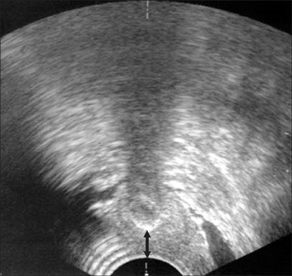
FIG. 1 Measurement of proximal urethrovaginal space by transvaginal ultrasonography (the arrow represents the distance between the anterior vaginal wall and the catheter balloon).

FIG. 2 Measurement of urethral length by use of a Foley catheter (the mark on the catheter represents the point of the urethral meatus and the proximal end of the balloon represents the point of the bladder neck, respectively).
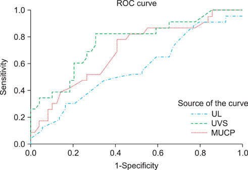
FIG. 3 Receiver operating characteristic (ROC) curve of urethral length (UL), proximal urethrovaginal space (UVS), and maximal urethral closure pressure (MUCP) for detrusor overactivity (AUC: 0.567, 0.763, and 0.694, respectively).
Reference
-
1. Seo JB, Lee JZ. The epidemiologic study of the urinary incontinence in community-dwelling women over 50 years old. Korean J Urol. 1999. 40:1525–1530.2. Brubaker L, Stoddard A, Richter H, Zimmern P, Moalli P, Kraus SR, et al. Mixed incontinence: comparing definitions in women having stress incontinence surgery. Neurourol Urodyn. 2009. 28:268–273.3. Abrams P, Cardozo L, Fall M, Griffiths D, Rosier P, Ulmsten U, et al. The standardisation of terminology of lower urinary tract function: report from the standardisation sub-committee of the international continence society. Neurourol Urodyn. 2002. 21:167–178.4. Simeonova Z, Milsom I, Kullendorff AM, Molander U, Bengtsson C. The prevalence of urinary incontinence and its influence on the quality of life in women from an urban Swedish population. Acta Obstet Gynecol Scand. 1999. 78:546–551.5. Dmochowski R, Staskin D. Mixed incontinence: definitions, outcomes, and interventions. Curr Opin Urol. 2005. 15:374–379.6. Kim JJ, Bae JH, Lee JG. Preoperative factors predicting the outcome of a midurethal sling operation for treating women with mixed incontinence. Korean J Urol. 2008. 49:1112–1118.7. Tunn R, Goldammer K, Neymeyer J, Gauruder-Burmester A, Hamm B, Beyersdorff D. MRI morphology of the levator ani muscle, endopelvic fascia, and urethra in women with stress urinary incontinence. Eur J Obstet Gynecol Reprod Biol. 2006. 126:239–245.8. Choi JD, Koh SB, Kim DY. Comparison of urethral length and anterior vaginal wall thickness between continent and incontinent women. Korean J Urol. 2009. 50:28–32.9. Jung SY, Fraser MO, Ozawa H, Yokoyama O, Yoshiyama M, De Groat WC, et al. Urethral afferent nerve activity affects the micturition reflex; Implication for the relationship between stress incontinence and detrusor instability. J Urol. 1999. 162:204–212.10. Schäfer W, Abrams P, Liao L, Mattiasson A, Pesce F, Spangberg A, et al. Good urodynamic practices: uroflowmetry, filling cystometry, and pressure-flow studies. Neurourol Urodyn. 2002. 21:261–274.11. Chaliha C, Khullar V. Mixed incontinence. Urology. 2004. 63:3 Suppl 1. 51–57.12. Anger JT, Rodríguez LV. Mixed incontinence: stressing about urge. Curr Urol Rep. 2004. 5:427–431.13. Yun HC, Lee JG. Is urodynamic evaluation necessary for women with stress urinary incontinence? Korean J Urol. 2002. 43:687–692.14. Bates CP, Loose H, Stanton SL. The objective study of incontinence after repair operations. Surg Gynecol Obstet. 1973. 136:17–22.15. Tunuguntla HS, Gousse AE. Female sexual dysfunction following vaginal surgery: a review. J Urol. 2006. 175:439–446.16. Barber MD, Visco AG, Wyman JF, Fantl JA, Bump RC. Continence Program for Women Research Group. Sexual function in women with urinary incontinence and pelvic organ prolapse. Obstet Gynecol. 2002. 99:281–289.17. Gravina GL, Brandetti F, Martini P, Carosa E, Di Stasi SM, Morano S, et al. Measurement of the thickness of the urethrovaginal space in women with or without vaginal orgasm. J Sex Med. 2008. 5:610–618.18. Ibe C, Simon JA. Vulvovaginal atrophy: current and future therapies (CME). J Sex Med. 2010. 7:1042–1050.19. Lynch C. Vaginal estrogen therapy for the treatment of atrophic vaginitis. J Womens Health (Larchmt). 2009. 18:1595–1606.20. Koo JM, Lee TY. Characteristic changes of cough urethral pressure profile in stress urinary incontinence. Korean J Urol. 1994. 35:665–670.21. Digesu GA, Robinson D, Cardozo L, Khullar V. Three-dimensional ultrasound of the urethral sphincter predicts continence surgery outcome. Neurourol Urodyn. 2009. 28:90–94.22. Kondo Y, Homma Y, Takahashi S, Kitamura T, Kawabe K. Transvaginal ultrasound of urethral sphincter at the mid urethra in continent and incontinent women. J Urol. 2001. 165:149–152.23. Major H, Culligan P, Heit M. Urethral sphincter morphology in women with detrusor instability. Obstet Gynecol. 2002. 99:63–68.24. Awad S, McGinnis R. Factors that influence the incidence of detrusor instability in women. J Urol. 1983. 130:114–115.25. Paick JS, Ku JH, Kim SW, Oh SJ, Son H, Shin JW. Tension-free vaginal tape procedure for the treatment of mixed urinary incontinence: significance of maximal urethral closure pressure. J Urol. 2004. 172:1001–1005.26. Woo JH, Hong SJ, Lee JB. The relationship of pressure-flow parameters and urethral pressure in female patients with lower urinary tract symptoms. Korean J Urol. 2009. 50:567–572.
- Full Text Links
-
- Actions
-
Cited
- CITED
-
- Close
- Share
-
- Similar articles
-
- Behcet's Disease with Urethrovaginal Fistula and Stress Urinary Incontinence
- Urodynamic Detrusor Overactivity in Patients with Overactive Bladder Symptoms
- The Relationship of Pressure-Flow Parameters and Urethral Pressure in Female Patients with Lower Urinary Tract Symptoms
- Comparison between Natural Filling Cystometry and Conventional Retrograde Filling Cystometry in Patients with Stroke
- Is Urodynamic Evaluation Necessary for Women with Stress Urinary Incontinence?
