Efficacy of Renal Artery Embolization using a Mixture of Histoacryl(R) and Lipiodol in a Rabbit Model
- Affiliations
-
- 1Department of Diagnostic Radiology, College of Medicine, The Catholic University of Korea, Seoul, Korea.
- 2Department of Diagnostic Urology, College of Medicine, The Catholic University of Korea, Seoul, Korea. urokhw@catholic.ac.kr
- KMID: 2061289
- DOI: http://doi.org/10.4111/kju.2007.48.9.903
Abstract
- PURPOSE
We wanted to evaluate the efficacy and computed tomography(CT) findings of renal artery embolization with using a mixture of Histoacryl(R) and lipiodol in rabbit depending on the mixture proportions and the temporal course.
MATERIALS AND METHODS
Eighteen rabbits were equally divided into two groups: group A received a 1:3 mixture and group B received a 1:5 mixture of Histoacryl(R) and lipiodol. We subdivided each group as follows: the 1-day group, the 10-day group and the 20-day group according to the elapsed days after embolization, respectively. As a result, the experimental groups were composed of six subgroups. Afterright renal artery embolizations, plain abdominal radiographs were obtained from all the rabbits. On the first day, the 10th day and the 20th day after embolization, abdominal CT was performed in each subgroup.
RESULTS
On the post-embolization radiographs, the embolic casts were formed only at the main or segmental renal arteries in 7 cases of group A. On the other hand, the embolic casts were formed at the entire arterial trees in two cases of group A and all the cases of group B. On the pre- contrast-enhanced CT scans, there were radiopaque densities of embolic casts, residual lipiodol flecks and calcifications in the embolized kidneys. On the contrast-enhanced CT scans, global perfusion defects of the kidneys were noted in 17 rabbits. The cortical rim signs were noted in all rabbits of the 10-day and 20-day groups, except for one rabbit.
CONCLUSIONS
The mixture of Histoacryl(R) and lipiodol is effective for renal artery embolization. The CT findings of the embolized kidneys are characteristic depending on the mixture proportions of the embolic agents and the temporal courses.
Keyword
MeSH Terms
Figure
-
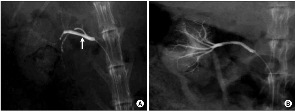
Fig. 1 Immediate post-embolization angiograms show a radio-opaque embolic cast of Histoacryl® and lipiodol in the main renal artery (arrow) in group A (A) and in the entire arterial trees of the kidney in group B (B).
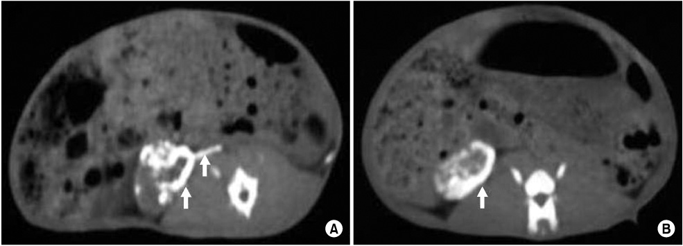
Fig. 2 After embolization, pre-contrast CT scans show linear and nodular radiopaque embolic casts and lipiodol flecks (arrows) in the main renal artery and kidney of group A (A), and circumferential lipiodol flecks (arrow) were seen in the kidney, mainly at the cortex of group B (B).
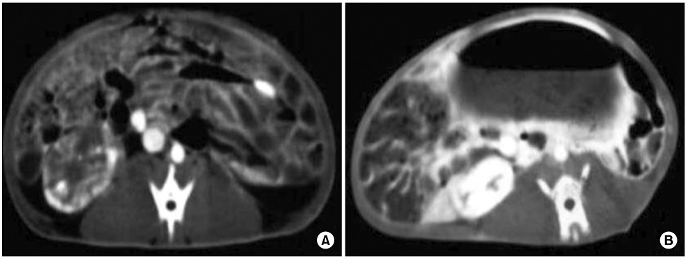
Fig. 3 After embolization, post-contrast CT scans show the complete absence of renal enhancement in groups A and B (A) with only one exception in group B-3 (B).
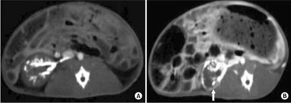
Fig. 4 After embolization, post-contrast CT scans show no cortical rim sign in group A-1 (A) and a cortical rim sign (arrow) is seen in group A-3 (B).
Reference
-
1. Lalli AF, Peterson N, Bookstein JJ. Roentgen-guided infarctions of kidneys and lungs. A potential therapeutic technic. Radiology. 1969. 93:434–435.2. Almgard LE, Fernstrom I, Haverling M, Ljungqvist A. Treatment of adenocarcinoma by embolic occlusion of the renal circulation. Br J Urol. 1973. 45:474–479.3. Papo J, Baratz M, Merimsky E. Infarction of renal tumors using isobutyl-2-cyanoacrylate and lipiodol. AJR Am J Roentgenol. 1981. 137:781–785.4. Wallace S, Chuang VP, Swanson D, Bracken B, Hersh EM, Ayala A, et al. Embolization of renal carcinoma. Radiology. 1981. 138:563–570.5. Moorhead JD, Fritzsche P, Hadley HL. Management of hemorrhage secondary to renal angiomyolipoma with selective arterial embolization. J Urol. 1977. 117:122–123.6. Ellman BA, Parkhill BJ, Curry TS, Marcus PB, Peters PC. Ablation of renal tumors with absolute ethanol: a new technique. Radiology. 1981. 141:619–626.7. Ekelund L, Jonsson N, Treugut H. Transcatheter obliteration of the renal artery by ethanol injection: experimental results. Cardiovasc Intervent Radiol. 1981. 4:1–7.8. Mazer MJ, Baltaxe HA, Wolf GL. Therapeutic embolization of the renal artery with Gianturco coil: limitations and technical pitfalls. Radiology. 1981. 138:37–46.9. Barth KH, Strandberg JD, White RI Jr. Long term follow-up of transcatheter embolization with autologous clot, oxycel and gelfoam in domestic swine. Invest Radiol. 1977. 12:273–280.10. Soehendra N, Nam VC, Grimm H, Kempeneers I. Endoscopic obliteration of large esophagogastric varices with bucrylate. Endoscopy. 1986. 18:25–26.11. Wikholm G, Lundqvist C, Svendsen P. Embolization of cerebral arteriovenous malformations: part I-technique, morphology and complications. Neurosurgery. 1996. 39:448–459.12. Giuliani L, Carmignani G, Belgrano E, Puppo P. Transcatheter arterial embolization in urological tumors: the use of isobutyl-2-cyanoacrylate. J Urol. 1979. 121:630–634.13. Gunther R, Schubert U, Bohl J, Georgi M, Marberger M. Transcatheter embolization of the kidney with butyl-2-cyanoacrylate: experimental and clinical results. Cardiovasc Radiol. 1978. 1:101–108.14. Kaisary AV, Williams G, Riddle PR. The role of preoperative embolization in renal cell carcinoma. J Urol. 1984. 131:641–646.15. Craven WM, Redmond PL, Kumpe DA, Durham JD, Wettlaufer JN. Planned delayed nephrectomy after ethanol embolization of renal carcinoma. J Urol. 1991. 146:704–708.16. Alvarez Gonzalez E, Pamplona M, Rodriguez A, Garcia-Hidalgo E, Nunez V, Leiva O. High flow priapism after blunt perineal trauma: resolution with bucryllate embolization. J Urol. 1994. 151:426–428.17. Peskircioglu L, Tekin MI, Boyvat F, Karabulut A, Ozkardes H. Embolization of the deep dorsal vein for the treatment of erectile impotence due to veno-occlusive dysfunction. J Urol. 2000. 163:472–475.18. Yok KY, Kum CK, Goh PM. Endoscopic hemostasis of upper gastrointestinal bleeding with histoacryl: last resort before surgery. Endoscopy. 1996. 28:256–258.19. Soehendra N, Nam VC, Grimm H, Kempeneers I. Endoscopic obliteration of large esophagogastric varices with bucrylate. Endoscopy. 1986. 18:25–26.20. Spiegel SM, Vinuela F, Goldwasser JM, Fox AJ, Pelz DM. Adjusting the polymerization time of isobutyl-2 cyanoacrylate. Am J Neuroradiol. 1986. 7:109–112.21. Suh DC, Kim HJ, Kim ST, Choi CG, Lee HK, Song HY, et al. Experimental evaluation of embolic effect: pure glue and glue-tungsten mixture. J Korean Radiol Soc. 1997. 37:963–968.22. Glazer GM, Francis IR, Brady TM, Teng SS. Computed tomography of renal infarction: clinical and experimental observations. AJR Am J Roentgenol. 1983. 140:721–727.23. Wong WS, Moss AA, Federle MP, Cochran ST, London SS. Renal infarction: CT diagnosis and correlation between CT findings and etiologies. Radiology. 1984. 150:201–205.24. Kamel IR, Berkowitz JF. Assessment of the cortical rim sign in posttraumatic renal infarction. J Comput Assist Tomogr. 1996. 20:803–806.
- Full Text Links
-
- Actions
-
Cited
- CITED
-
- Close
- Share
-
- Similar articles
-
- Experimental Embolization Using Histoacryl Blue (N-butyl 2-cyanoacrylate) in Pig Rete Mirabile
- Safety of Superselective Transcatheter Arterial Chemoembolization through Cystic Artery for Treatment of Hepatocellular Carcinoma
- A Case of Portal and Splenic Vein Thrombosis Developed by Complication of Histoacryl Injection Therapy in Gastric Varix
- The Arterial Embolization with Glue-Lipiodol Mixture in Patients with Hemoptysis
- A microangiographic study on renal artery embolization
