Focal Myositis around Hip Joint: 3 Cases Report
- Affiliations
-
- 1Department of Orthopedic Surgery, College of Medicine, Konyang University, Daejeon, Korea. ajouos@hanmail.net
- KMID: 2054186
- DOI: http://doi.org/10.5371/hp.2014.26.3.198
Abstract
- Focal myositis, a benign myositis which mostly occurs at lower extremity, is a disease that is spontaneously improved by conservative treatments such as bed rest and administration of nonsteroidal anti-inflammatory drug. Focal myositis is known to occur mostly at lower extremity, but we could not find a report of occurrence around hip. Therefore, authors attempt to report clinical progression along with the literature review.
Keyword
Figure
-

Fig. 1 Hip magnetic resonance imaging. (A) T2 fat suppression image showed increased ill-defined signal intensity on gluteus medius muscle. (B) T1 contrast enhanced fat suppression image showed enhancement on gluteus medius muscle. (C) After 3 months follow up, T2 image showed recovery of previous abnormal finding.
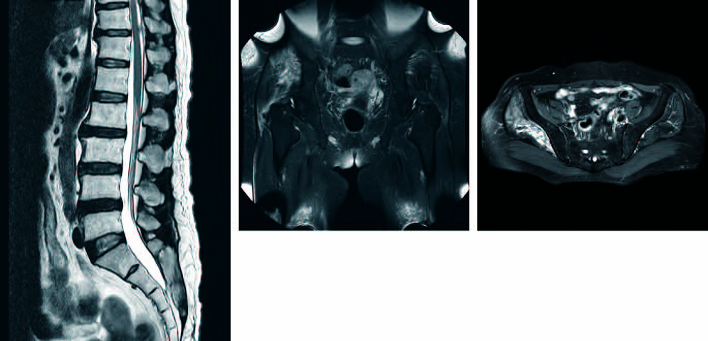
Fig. 2 Spine and hip magnetic resonance imaging (MRI). (A) In lumbar spine MRI, there is no abnormal finding that explain patient's pain. (B) T2 fat suppression image showed increased ill-defined signal intensity on gluteus medius and minimus muscles. (C) T1 contrast enhanced fat suppression image showed A enhancement on gluteus medius and minimus muscles.

Fig. 3 Hip magnetic resonance imaging. (A) T2 fat suppression image showed ill-defined signal change on left iliopsoas muscle. (B) T1 contrast enhanced fat suppression image showed contrast enhancement on left iliacus muscle.
Reference
-
1. Heffner RR Jr, Armbrustmacher VW, Earle KM. Focal myositis. Cancer. 1977; 40:301–306.
Article2. Auerbach A, Fanburg-Smith JC, Wang G, Rushing EJ. Focal myositis: a clinicopathologic study of 115 cases of an intramuscular mass-like reactive process. Am J Surg Pathol. 2009; 33:1016–1024.3. Llauger J, Bagué S, Palmer J, Matías-Guiu X, San Román L, Doncel A. Focal myositis of the thigh: unusual MR pattern. Skeletal Radiol. 2002; 31:307–310.
Article4. Prop S, van Vuurden D, van der Kuip M, van der Voorn JP, Plötz FB. A boy with cervical focal myositis. Ned Tijdschr Geneeskd. 2014; 158:A6935.5. Heffner RR Jr, Barron SA. Polymyositis beginning as a focal process. Arch Neurol. 1981; 38:439–442.
Article6. Flaisler F, Blin D, Asencio G, Lopez FM, Combe B. Focal myositis: a localized form of polymyositis? J Rheumatol. 1993; 20:1414–1416.7. Brown P, Doyle DV, Evans MD. Localized nodular myositis as the first manifestation of polymyositis. Br J Rheumatol. 1989; 28:84.
Article8. Garcia-Consuegra J, Morales C, Gonzalez J, Merino R. Relapsing focal myositis: a case report. Clin Exp Rheumatol. 1995; 13:395–397.9. Misu T, Tateyama M, Nakashima I, Shiga Y, Fujihara K, Itoyama Y. Relapsing focal myositis: the localization detected by gallium citrate Ga 67 scintigraphy. Arch Neurol. 2005; 62:1930–1931.10. Jun J, Im S, Park JH, Yoo SH, Park GY. Focal myositis of unilateral leg. Ann Rehabil Med. 2011; 35:944–948.
Article
