Study on branching pattern of aortic arch in Indian
- Affiliations
-
- 1Department of Anatomy, Government Medical College, Nagpur, Maharashtra, India. dr.sumitpatil1122@gmail.com
- KMID: 2046748
- DOI: http://doi.org/10.5115/acb.2012.45.3.203
Abstract
- Knowledge of the branching pattern of aortic arch is important during supra-aortic angiography, aortic instrumentation, thoracic and neck surgery. The purpose of this study is to describe different branching pattern of arch of aorta in Indian subjects, in order to offer useful data to anatomists, radiologists, vascular surgeons while relating it to the embryological basis. Seventy five arches of adult Indian cadavers were exposed and their branches examined during cadaveric dissection in the Department of Anatomy, Government Medical College, Nagpur. The usual three-branched aortic arch was found in 58 cadavers (77.3%); the 11 (14.66%) remaining aortic arch showed only two branches, out of which one was a common trunk, which incorporated the brachiocephalic trunk and left common carotid and other left subclavian artery and 6 (8%) aortic arches showed direct arch origin of the left vertebral artery. Although the variations are usually asymptomatic, they may cause dyspnoea, dysphasia, intermittent claudication, misinterpretation of radiological examinations and complications during neck and thorax surgery. Knowledge of different patterns of arch of aorta is critical when invading the arch of aorta and its branches by instruments, as all these areas are delicate.
Keyword
MeSH Terms
Figure
-
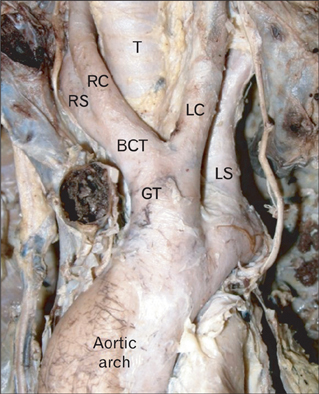
Fig. 1 Showing type II A aortic arch. BCT, brachiocephalic trunk; GT, great trunk; LC, left common carotid; LS, left subclavian; RC, right common carotid; RS, right subclavian; T, trachea.
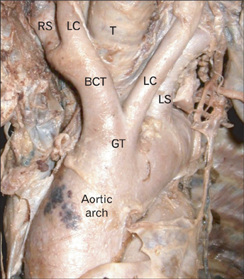
Fig. 2 Showing type II B aortic arch. BCT, brachiocephalic trunk; GT, great trunk; LC, left common carotid; LS, left subclavian; RS, right subclavian; T, trachea.
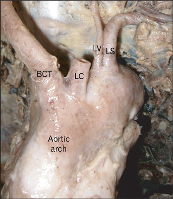
Fig. 3 Showing type III aortic arch. BCT, brachiocephalic trunk; LC, left common carotid; LS, left subclavian; LV, left vertebral artery.

Fig. 4 Radiological images of aortic arch showing (A) type I, (B) type II, and (C) type III branching pattern of aortic arch. BCT, brachiocephalic trunk; GT, great trunk; LC, left common carotid; LS, left subclavian; LV, left vertebral artery; RS, right subclavian.
Cited by 1 articles
-
Right and left common carotid arteries arising from the branchiocephalic, a rare variation of the aortic arch
Eleni Panagouli, Gregory Tsoucalas, Theodoros Papaioannou, Aliki Fiska, Dionysios Venieratos, Panagiotis Skandalakis
Anat Cell Biol. 2018;51(3):215-217. doi: 10.5115/acb.2018.51.3.215.
Reference
-
1. Williams PL, Warwick R, Dyson M, Bannister LH. Gray's anatomy. 1989. 37th ed. Edinburgh: Churchill Livingstone;733–734.2. Luisada A. Cardiology. 1963. Vol. 1. New York: McGraw-Hill;68.3. Wells TR, Landing BH, Shankle WR. Syndromal associations of common origin of the carotid arteries. Pediatr Pathol. 1993. 13:203–212.4. Williams GD, Edmonds HW. Variations in the arrangement of the branches arising from the aortic arch in American whites and Negroes. Anat Rec. 1935. 62:139–146.5. Natsis KI, Tsitouridis IA, Didagelos MV, Fillipidis AA, Vlasis KG, Tsikaras PD. Anatomical variations in the branches of the human aortic arch in 633 angiographies: clinical significance and literature review. Surg Radiol Anat. 2009. 31:319–323.6. Shiva Kumar GL, Pamidi N, Somayaji SN, Nayak S, Vollala VR. Anomalous branching pattern of the aortic arch and its clinical applications. Singapore Med J. 2010. 51:e182–e183.7. Moore KL, Persaud TV. The developing human: clinically oriented embryology. 2008. 8th ed. Philadelphia: Saunders Elsevier;305–306. 316–325.8. Sadler TW. Langman's medical embryology. 2006. 10th ed. Philadelphia: Lippincott Williams & Wilkins;173–175. 180–185.9. Nelson ML, Sparks CD. Unusual aortic arch variation: distal origin of common carotid arteries. Clin Anat. 2001. 14:62–65.10. Satyapal KS, Singaram S, Partab P, Kalideen JM, Robbs JV. Aortic arch branch variations: case report and arteriographic analysis. S Afr J Surg. 2003. 41:48–50.11. Adachi B. Das Arterien system der Japaner. 1928. Vol. 1. Kyoto: Verlag der Kaiserlich-Japanischen Universitat, Kenyusha Press;29–41.12. Anson BJ, McVay CB. Surgical anatomy. 1971. 5th ed. Philadelphia, London, Toronto: W.B. Saunders;408–412.13. Paraskevas G, Agios P, Stavrakas M, Stoltidou A, Tzaveas A. Left common carotid artery arising from the brachiocephalic trunk: a case report. Cases J. 2008. 1:83.
- Full Text Links
-
- Actions
-
Cited
- CITED
-
- Close
- Share
-
- Similar articles
-
- Morphology of the aortic arch branching pattern in raccoon dogs (Nyctereutes procyonoides, Gray, 1834)
- Complete Vascular Ring Caused by Kommerell's Diverticulum and Right Aortic Arch with Mirror Image Branching
- A Case of the Right Aortic Arch with Mirror-image Branching and a Left Ligamentum Arteriosum Forming a Vascular Ring
- A Case of Right Sided Aortic Arch Combined with Atrial Septal Defect
- Hybrid Procedure for Aortic Arch Repair: Arch Vessels Debranching with Supraaortic Revascularization Followed by Endovascular Aortic Stent Grafting
