Two Consecutive Levels of Unilateral Cervical Spondylolysis on Opposite Sides
- Affiliations
-
- 1Department of Radiology, Inje University College of Medicine, Haeundae Paik Hospital, Busan, Korea. bluesingirl@naver.com
- 2Department of Radiology, Myongji Hospital, Seonam University College of Medicine, Goyang, Korea.
- 3Department of Radiology, Dongguk University College of Medicine, Ilsan Hospital, Goyang, Korea.
- KMID: 2043609
- DOI: http://doi.org/10.3348/jksr.2015.73.3.181
Abstract
- Cervical spondylolysis, with or without spondylolisthesis, is a rare condition defined as a corticated cleft between the superior and inferior articular facets of the articular pillar. The defect occurs predominantly at C6, and is usually bilateral in up to two-thirds of cases. Multilevel involvement is uncommon, however, to date, no case of two consecutive levels of unilateral cervical spondylolysis on opposite sides has been reported. Here, we report a rare case of a patient affected by two consecutive levels of unilateral cervical spondylolysis at C5 and C6 on opposite sides in a 19-year-old male complaining of neck pain.
Figure
-

Fig. 1 Plain radiographs of the cervical spine. A. Anteroposterior radiograph showing spina bifida of C5 (arrow). B. Lateral radiograph in flexion showing well-defined spondylolytic defects that divide the articular pillar of C5 (arrow 1) and C6 (arrow 2) vertebra into two fragments. Spina bifida (arrow 3) of C5 is also revealed.

Fig. 2 CT scans of the cervical spine. A. Axial CT image showing a smoothly-marginated defect in the right articular pillar of C5 (arrow). B. Axial CT image showing a well-corticated defect in the left articular pillar of C6 with irregular-shaped ossification (arrow) between the ventral (V) and dorsal fragment (D). C. Axial CT image revealing spina bifida (arrow 1) and right dysplastic lamina (arrow 2) of C5. D. Axial CT image showing left dysplastic lamina of C6 (arrow 1), articulation (arrow 2) between the dorsal fragment of the C6 articular pillar and C7, and ossification (arrow 3) within the defect in the left articular pillar of C6. E. Reformatted sagittal CT image on the right side revealing a defect (arrow) in the right articular pillar of C5. The ventral fragment (V) of the articular pillar of C5 articulates with the facet of C4, and the dorsal fragment (D) articulates with the facet of C6. F. Reformatted sagittal CT image on the left side showing ossifications (arrows) in the defect in the C6 left articular pillar. The ventral (V) and dorsal (D) fragments articulate with the facets of C5 and C7, respectively. G, H. Three-dimensional volume rendering-CT images showing dysplastic laminae (L), corticated clefts (arrows) dividing the articular pillars into ventral (V) and dorsal (D) fragments.
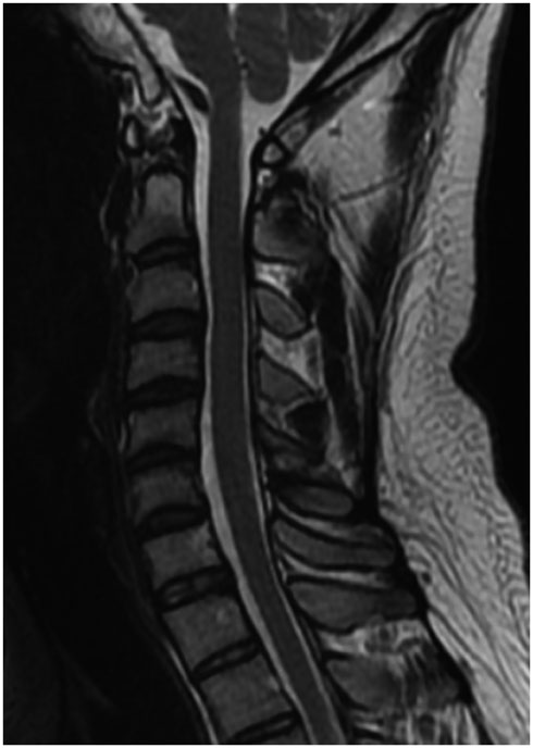
Fig. 3 MR image of the cervical spine. Sagittal T2-weighted MR image showing no evidence of spinal cord compression or signal abnormality, discoligamentous structure injury or bone marrow edema.
Reference
-
1. Forsberg DA, Martinez S, Vogler JB 3rd, Wiener MD. Cervical spondylolysis: imaging findings in 12 patients. AJR Am J Roentgenol. 1990; 154:751–755.2. Redla S, Sikdar T, Saifuddin A, Taylor BA. Imaging features of cervical spondylolysis--with emphasis on MR appearances. Clin Radiol. 1999; 54:815–820.3. Schwartz JM. Case 36: bilateral cervical spondylolysis of C6. Radiology. 2001; 220:191–194.4. Paik NC. Bilateral cervical spondylolysis of C7. Spine J. 2010; 10:e10–e13.5. Mofidi A, Tansey C, Mahapatra SR, Mirza HA, Eisenstein SM. Cervical spondylolysis, radiologic pointers of stability and acute traumatic as opposed to chronic spondylolysis. J Spinal Disord Tech. 2007; 20:473–479.6. Ahn PG, Yoon DH, Shin HC, Kim KN, Yi S, Lee DY, et al. Cervical spondylolysis: three cases and a review of the current literature. Spine (Phila Pa 1976). 2010; 35:E80–EE8.7. Kushare IV, Colo D, Kadhim M, Dormans JP. Bilateral C6 spondylolysis with spondylolisthesis in 3 adolescent siblings. J Pediatr Orthop. 2014; 34:e40–e43.8. Morvan G, Busson J, Frot B, Nahum H. [Cervical spondylolysis. 7 cases. Review of the literature]. J Radiol. 1984; 65:259–266.9. Karasick S, Karasick D, Wechsler RJ. Unilateral spondylolysis of the cervical spine. Skeletal Radiol. 1983; 9:259–261.10. Hinton MA, Harris MB, King AG. Cervical spondylolysis. Report of two cases. Spine (Phila Pa 1976). 1993; 18:1369–1372.
- Full Text Links
-
- Actions
-
Cited
- CITED
-
- Close
- Share
-
- Similar articles
-
- Cervical Spondylolysis in Child with Four Levels of Simultaneous Involvement: A Case Report
- Utilization of Three-dimensional Reconstruction Computed Tomography as a Diagnostic Tool for Adult Unilateral Primary Cervical Spondylolysis: Report of Two Cases
- Cervical Spondylolysis: Report of Two Cases
- Characteristics of Lumbar Spondylolysis in Adolescent Baseball Players: Relationship between the Laterality of Lumbar Spondylolysis and the Throwing or Batting Side
- Sequential Proximal Adjacent Spondylolysis by Pars Interarticularis Fracture in Elite Soccer Player
