Desmoplastic Fibroma of Bone: Case Report
- Affiliations
-
- 1Department of Diagnostic Radiology, Dong-A University College of Medicine, Korea. hdhdoc@naver.com
- 2Department of Pathology, Dong-A University College of Medicine, Korea.
- KMID: 2040818
- DOI: http://doi.org/10.3348/jksr.2011.64.5.491
Abstract
- Desmoplastic fibroma of bone is a rare benign primary bone tumor that histologically resembles the extra-abdominal desmoid tumor of soft tissues. It is a nonmetastasizing, but locally aggressive tumor that is similar to a desmoid tumor of the soft tissues, and so it is considered "semimalignant". According to a previous report on a series of bone tumors, the incidence rate of desmoplastic fibroma was 0.1-0.3%. Its rarity results in radiologists having a tendency of overlooking the possibility of desmoplastic fibroma of bone during the imaging readings. We report on the imaging findings of desmoplastic fibroma of bone with a review of the relevant literature.
Figure
-
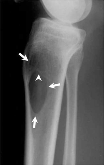
Fig. 1 The plain lateral and anteroposterior (not shown) radiographs of the proximal right tibia in a 36-year-old woman show a well marginated, osteolytic lesion (arrows) with internal trabeculation (arrow head) within the proximal tibial metadiaphysis. There is no evidence of matrix mineralization or cortical destruction.

Fig. 2 The coronal T1-weighted images (A) show an isointense lesion compared to the adjacent normal muscle within the proximal tibial metadiaphysis with cortical destruction and a soft tissue extension. The axial T2-weighted image (B) at a same level shows a hyperintense lesion in the medullary area (asterisk), which is hypointense to isointense relative to the peripheral area (arrow head). Note that the area of T2 low signal is more extensive than the hyperintense medullary area. The axial T1-wegihted image after gadolinium contrast administration (C) shows heterogeneous enhancement of the lesion. The hyperintense area on the T2-weighted image (asterisk) is more intensely enhanced than the area with T2 low signal (arrow head). Microscopic examination (D) shows monotonous spindle shaped fibroblasts with slender nuclei interspersed in the hyaline stroma of thick, wavy bands of collagen fiber (H & E, × 200).
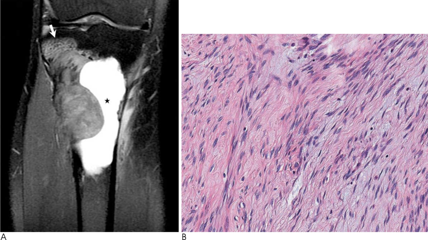
Fig. 3 MRI is performed to determine whether there is tumor recurrence. The coronal proton density-weighted fat suppressed image (A) shows a hyperintense lesion with foci of T2 low signal within the lateral proximal tibial metadiaphysis. The cavity of fluid signal intensity in the medulla (asterisk) seems to be the result of previous curettage. In contrast to the previous lesion in Fig. 2A, this lesion extends into the epiphysis (arrow). Note that the overall signal intensity of the lesion is higher than that of the previous lesion in Fig. 2B. The histologic specimen (B) shows similar characteristics as those of the previous specimen depicted in Fig. 2D. This specimen has more cellular areas and a lesser amount of collagenous matrix when compared with the previous specimen depicted in Fig. 2D.
Reference
-
1. Jaffe HL. Tumors and tumorous conditions of the bones and joints. Philadelphia: Lea & Febiger;1958. p. 298–303.2. Fornasico V, Pritzker KPH, Bridge JA. Pathology and genetics of tumours of soft tissue and bone. In : Fletcher CDM, Unni KK, Merten SF, editors. Pathology and genetics of tumours of soft tissue and bone. Lyon: IARC Press;2002. p. 288.3. Inwards CY, Unni KK, Beabout JW, Sim FH. Desmoplastic fibroma of bone. Cancer. 1991; 68:1978–1983.4. Mirra JM, Picci P, Gold RH. Bone tumors: clinical, radiologic, and pathologic correlations. Philadelphia: Lea & Febiger;1989. p. 735–747.5. Crim JR, Gold RH, Mirra JM, Eckardt JJ, Bassett LW. Desmoplastic fibroma of bone: radiographic analysis. Radiology. 1989; 172:827–832.6. Taconis WK, Schutte HE, van der Heul RO. Desmoplastic fibroma of bone: a reported of 18 cases. Skeletal Radiol. 1994; 23:283–288.7. Bohm P, Krober S, Greschniok A, Laniado M, Kaiserling E. Desmoplastic fibroma of the bone. A reported of two patients, review of the literature, and therapeutic implications. Cancer. 1996; 78:1011–1023.8. Frick MA, Sundaram M, Unni KK, Inwards CY, Fabbri N, Tretani F, et al. Imaging findings in desmoplastic fibroma of bone: distinctive T2 chracteristic. AJR Am J Roentgenol. 2005; 184:1762–1767.9. Vanhoenacker FM, Hauben E, De Beuckeleer LH, Willemen D, Van Marck E, De Schepper AM. Desmoplastic fibroma of bone: MRI features. Skeletal Radiol. 2000; 29:171–175.10. Sundaram M, McGuire MH, Schajowicz F. Soft-tissue masses: histologic basis for decreased signal (short T2) on T2-weighted MR images. AJR Am J Roentgenol. 1987; 148:1247–1767.
