J Korean Surg Soc.
2010 Dec;79(Suppl 1):S41-S44. 10.4174/jkss.2010.79.Suppl1.S41.
Intussusception in an Adult due to Inverted Meckel's Diverticulum with Ectopic Pancreatic Tissue
- Affiliations
-
- 1Department of Surgery, St. Vincent's Hospital, College of Medicine, The Catholic University of Korea, Suwon, Korea. hj@catholic.ac.kr
- 2Department of Pathology, St. Vincent's Hospital, College of Medicine, The Catholic University of Korea, Suwon, Korea.
- 3Department of Radiology, St. Vincent's Hospital, College of Medicine, The Catholic University of Korea, Suwon, Korea.
- KMID: 2040551
- DOI: http://doi.org/10.4174/jkss.2010.79.Suppl1.S41
Abstract
- Adult intussusception is rare involving of only 1% of the causes of bowel obstruction. We report a case of a 39-year-old female with intussusceptions due to inverted Meckel's diverticulum. She visited our hospital for diffuse abdominal pain during 1 week and aggravated abdominal pain for 2 days. Vital signs were stable, and there was periumbilical tenderness. She had no history of abdominal operation. CT scan showed a 3.7x2.1 cm of fatty mass with focal intussusception in the distal ileum. When the emergency operation was performed, the patient was found to be suffering from ileocolic intussusception. A manual reduction of intussusception showed inverted Meckel's deverticulum at 65 cm proximal to the ileocecal valve, and the segmental resection of small bowel including a Meckel's diverticulum was performed. Pathologic examination revealed a Meckel's diverticulum containing a 0.6x0.6 cm sized aberrant pancreas.
Keyword
MeSH Terms
Figure
-
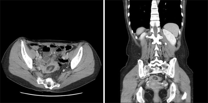
Fig. 1 CT scan shows fatty mass with focal intussusception in the distal ileum.
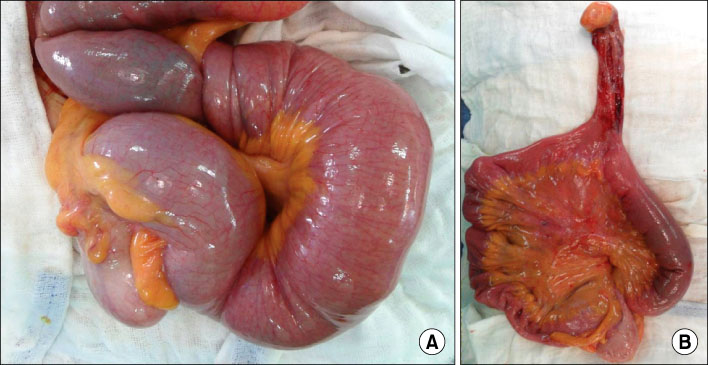
Fig. 2 Operative findings. (A) An ileoileal intussusception is noted. (B) After manual reduction of the intussusception, Meckel's diverticulum is found with a yellow colored soft mass on the tip.
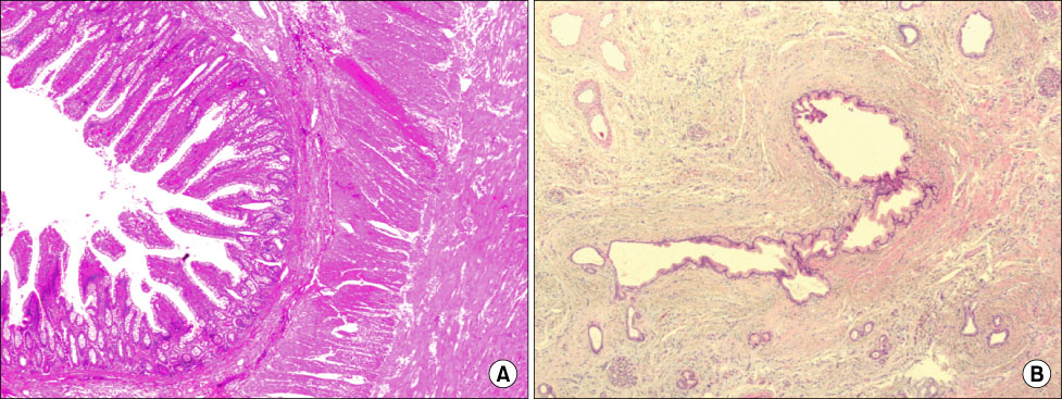
Fig. 3 Histologic findings. (A) The mucosal layer is composed of gastric, colonic or small intestinal mucosa. And the proper muscle layer is also found (H&E stain, ×40). (B) In the subserosal area, pancreatic tissue is noted forming round aggregate approximately 1.0×1.0 cm in size. The pancreatic tissue is composed of ductal epithelium and scattered clusters of endocrine cells in the fibrotic background (H&E stain, ×100).
Reference
-
1. Azar T, Berger DL. Adult intussusception. Ann Surg. 1997. 226:134–138.2. Stubenbord WT, Thorbjarnarson B. Intussusception in adults. Ann Surg. 1970. 172:306–310.3. Brayton D, Norris WJ. Intussusception in adults. Am J Surg. 1954. 88:32–43.4. Ymaguchi M, Takeuchi S, Awazu S. Meckel's diverticulum. Investigation of 600 patients in Japanese literature. Am J Surg. 1978. 136:247–249.5. Schwartz MJ, Lewis JH. Meckel's diverticulum: pitfalls in scintigraphic detection in the adult. Am J Gastroenterol. 1984. 79:611–618.6. Mackey WC, Dineen P. A fifty year experience with Meckel's diverticulum. Surg Gynecol Obstet. 1983. 156:56–64.7. Park JJ, Wolff BG, Tollefson MK, Walsh EE, Larson DR. Meckel diverticulum: the Mayo Clinic experience with 1476 patients (1950-2002). Ann Surg. 2005. 241:529–533.8. Pantongrag-Brown L, Levine MS, Elsayed AM, Buetow PC, Agrons GA, Buck JL. Inverted Meckel diverticulum: clinical, radiologic, and pathologic findings. Radiology. 1996. 199:693–696.9. Daneman A, Myers M, Shuckett B, Alton DJ. Sonographic appearances of inverted Meckel diverticulum with intussusception. Pediatr Radiol. 1997. 27:295–298.10. Hamada T, Ishida O, Yasutomi M. Inverted Meckel diverticulum with intussusception: demonstration by CT. J Comput Assist Tomogr. 1996. 20:287–289.
- Full Text Links
-
- Actions
-
Cited
- CITED
-
- Close
- Share
-
- Similar articles
-
- Intussusception due to Inverted Meckel Diverticulum with Ectopic Pancreas: A Case Report
- A Case of Intussusception Caused by Meckel's Diverticulum with Heterotopic Pancreatic and Gastric Tissues
- A Case of Chronic Intussusception Induced by Meckel`s Diverticulum in Adult
- Synchronous ileal inflammatory fibroid polyp and Meckel’s diverticulum found during laparoscopic surgery for adult intussusception
- A Case of Massive Hematochezia from a Meckel's Diverticulum without Ectopic Mucosa
