J Korean Neurosurg Soc.
2014 Oct;56(4):330-333. 10.3340/jkns.2014.56.4.330.
The Clinical Experience of Computed Tomographic-Guided Navigation System in C1-2 Spine Instrumentation Surgery
- Affiliations
-
- 1Department of Neurosurgery, Bucheon St. Mary's Hospital, The Catholic University of Korea College of Medicine, Bucheon, Korea. kimsd@catholic.ac.kr
- 2Department of Neurosurgery, Bonedream Hospital, Bucheon, Korea.
- KMID: 2018084
- DOI: http://doi.org/10.3340/jkns.2014.56.4.330
Abstract
OBJECTIVE
To identify the accuracy and efficiency of the computed tomographic (CT)-based navigation system on upper cervical instrumentation, particularly C1 lateral mass and C2 pedicle screw fixation compared to previous reports.
METHODS
Between May 2005 and March 2014, 25 patients underwent upper cervical instrumentation via a CT-based navigation system. Seven patients were excluded, while 18 patients were involved. There were 13 males and five females; resulting in four degenerative cervical diseases and 14 trauma cases. A CT-based navigation system and lateral fluoroscopy were used during the screw instrumentation procedure. Among the 58 screws inserted as C1-2 screws fixation, their precise positions were evaluated by postoperative CT scans and classified into three categories : in-pedicle, non-critical breach, and critical breach.
RESULTS
Postoperatively, the precise positions of the C1-2 screws fixation were 81.1% (47/58), and 8.6% (5/58) were of non-critical breach, while 10.3% (6/58) were of critical breach. Most (5/6, 83.3%) of the critical breaches and all of non-critical breaches were observed in the C2 pedicle screws and there was only one case of a critical breach among the C1 lateral mass screws. There were three complications (two vertebral artery occlusions and a deep wound infection), but no postoperative instrument-related neurological deteriorations were seen, even in the critical breach cases.
CONCLUSION
Although CT-based navigation systems can result in a more precise procedure, there are still some problems at the upper cervical spine levels, where the anatomy is highly variable. Even though there were no catastrophic complications, more experience are needed for safer procedure.
Keyword
MeSH Terms
Figure
-
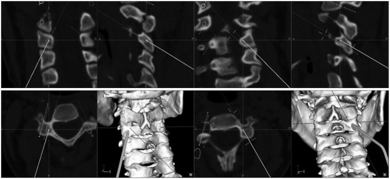
Fig. 1 Case 7, M/24, C1-2-3 posterior fixation. Simulation of pedicle screw insertion and preoperative screw trajectories can be obtained using a navigation system (Navigation cart II).
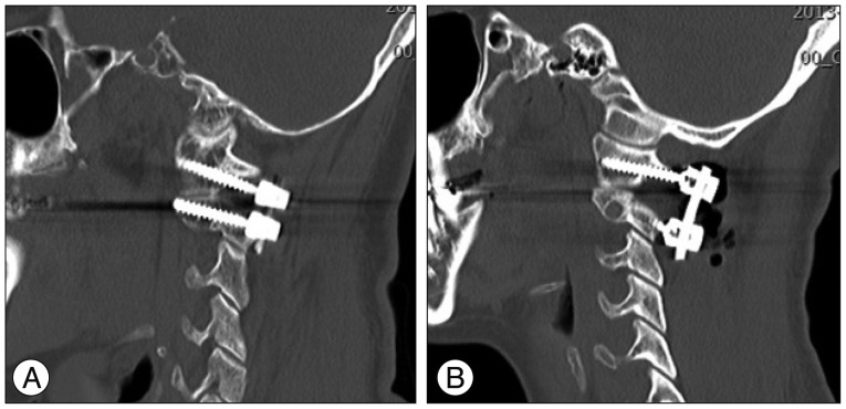
Fig. 2 A : Case 9, M/45, C1-2 posterior fixation via a typical C1-lateral mass screw insertion. B : Case 16, F/27, C1-2 posterior fixation via a modified C1-lateral mass screw with partial removal of inferior part of C1 posterior arch.
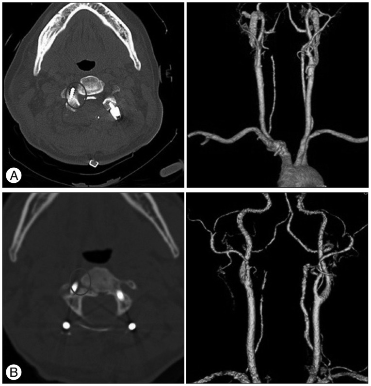
Fig. 3 A : Case 1, F/57, C2-3-4 posterior fixation. B : Case 15, M/48, C1-2-3 posterior fixation. Both cases had right side C2-pedicle screw violations in more than 50% (circles), but retrograde arterial flow from the opposite vertebral arteries are seen in the CT-angiogram.
Reference
-
1. Aryan HE, Newman CB, Nottmeier EW, Acosta FL Jr, Wang VY, Ames CP. Stabilization of the atlantoaxial complex via C-1 lateral mass and C-2 pedicle screw fixation in a multicenter clinical experience in 102 patients : modification of the Harms and Goel techniques. J Neurosurg Spine. 2008; 8:222–229. PMID: 18312073.
Article2. Gelalis ID, Paschos NK, Pakos EE, Politis AN, Arnaoutoglou CM, Karageorgos AC, et al. Accuracy of pedicle screw placement : a systematic review of prospective in vivo studies comparing free hand, fluoroscopy guidance and navigation techniques. Eur Spine J. 2012; 21:247–255. PMID: 21901328.
Article3. Goel A, Laheri V. Plate and screw fixation for atlanto-axial subluxation. Acta Neurochir (Wien). 1994; 129:47–53. PMID: 7998495.
Article4. Guppy KH, Chakrabarti I, Banerjee A. The use of intraoperative navigation for complex upper cervical spine surgery. Neurosurg Focus. 2014; 36:E5. PMID: 24580006.
Article5. Harms J, Melcher RP. Posterior C1-C2 fusion with polyaxial screw and rod fixation. Spine (Phila Pa 1976). 2001; 26:2467–2471. PMID: 11707712.
Article6. Hecht AC, Koehler SM, Laudone JC, Jenkins A, Qureshi S. Is intraoperative CT of posterior cervical spine instrumentation cost-effective and does it reduce complications? Clin Orthop Relat Res. 2011; 469:1035–1041. PMID: 20922584.
Article7. Ishikawa Y, Kanemura T, Yoshida G, Ito Z, Muramoto A, Ohno S. Clinical accuracy of three-dimensional fluoroscopy-based computer-assisted cervical pedicle screw placement : a retrospective comparative study of conventional versus computer-assisted cervical pedicle screw placement. J Neurosurg Spine. 2010; 13:606–611. PMID: 21039152.
Article8. Kim JH, Kwak DS, Han SH, Cho SM, You SH, Kim MK. Anatomic consideration of the C1 laminar arch for lateral mass screw fixation via C1 lateral lamina : a landmark between the lateral and posterior lamina of the C1. J Korean Neurosurg Soc. 2013; 54:25–29. PMID: 24044076.
Article9. Kotani Y, Abumi K, Ito M, Minami A. Improved accuracy of computer-assisted cervical pedicle screw insertion. J Neurosurg. 2003; 99(3 Suppl):257–263. PMID: 14563142.
Article10. Ludwig SC, Kramer DL, Balderston RA, Vaccaro AR, Foley KF, Albert TJ. Placement of pedicle screws in the human cadaveric cervical spine : comparative accuracy of three techniques. Spine (Phila Pa 1976). 2000; 25:1655–1667. PMID: 10870141.11. Moses ZB, Mayer RR, Strickland BA, Kretzer RM, Wolinsky JP, Gokaslan ZL, et al. Neuronavigation in minimally invasive spine surgery. Neurosurg Focus. 2013; 35:E12. PMID: 23905950.
Article12. Rampersaud YR, Pik JH, Salonen D, Farooq S. Clinical accuracy of fluoroscopic computer-assisted pedicle screw fixation : a CT analysis. Spine (Phila Pa 1976). 2005; 30:E183–E190. PMID: 15803068.13. Richter M, Cakir B, Schmidt R. Cervical pedicle screws : conventional versus computer-assisted placement of cannulated screws. Spine (Phila Pa 1976). 2005; 30:2280–2287. PMID: 16227890.
Article
- Full Text Links
-
- Actions
-
Cited
- CITED
-
- Close
- Share
-
- Similar articles
-
- Atlantoaxial Stabilization Using C1 Lateral Mass and C2 Pedicle/Translaminar Screw Fixation by Intraoperative C1- and C2-Direct-Captured Navigation with Preoperative Computed Tomography Images
- Comparative Analysis of Anterior Cervical Discectomy and Fusion at a Single Level: Free-Hand Versus Navigation-Guided Approaches
- Accuracy and Safety in Pedicle Screw Placement in the Thoracic and Lumbar Spines : Comparison Study between Conventional C-Arm Fluoroscopy and Navigation Coupled with O-Arm(R) Guided Methods
- Comparison of an Accelerometer-Based Portable Navigation System, Patient-Specific Instrumentation, and Conventional Instrumentation for Femoral Alignment in Total Knee Arthroplasty
- Comparative Analysis of Surgical Outcomes of C1–2 Fusion Spine Surgery between Intraoperative Computed Tomography Image Based Navigation-Guided Operation and Fluoroscopy-Guided Operation