Horizontal ridge expansion and implant placement using screws: a report of two cases
- Affiliations
-
- 1Department of Oral and Maxillofacial Surgery, Section of Dentistry, Seoul National University Bundang Hospital, Seongnam, Korea.
- 2Department of Dentistry and Dental Research Institute, School of Dentistry, Seoul National University, Seoul, Korea.
- 3Department of Oral and Maxillofacial Surgery, School of Dentistry, Chosun University, Gwangju, Korea. sgckim@chosun.ac.kr
- KMID: 2005453
- DOI: http://doi.org/10.5125/jkaoms.2014.40.5.233
Abstract
- Implants are typically placed after performing ridge expansion by inserting screws of gradually increasing thickness and good clinical outcomes are often obtained. We placed 11 implants in 6 patients, and one implant failed during osseointegration but it was replaced immediately after removal and successful prosthetic treatments were completed. During these surgeries, buccal cortical plate complete fractures do not occur. Inserting screws for ridge expansion is a successful and predictable technique for implant placement in narrow alveolar bone.
MeSH Terms
Figure
-
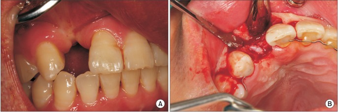
Fig. 1 Clinical view before implant placement. A. Preoperative intraoral photograph. B. Mucoperiosteal flap was elevated. Narrow alveolar ridge is observed.
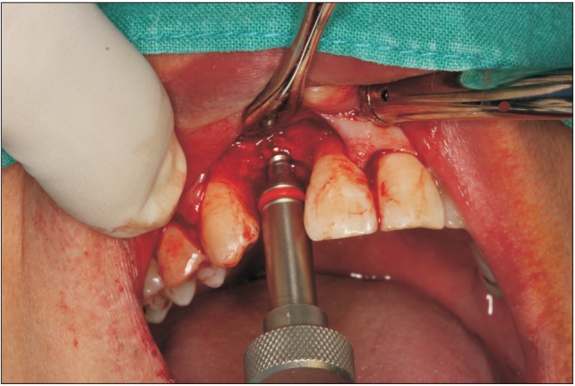
Fig. 2 Expansion of buccal plate achieved by splitting the ridge with bone expander in SplitMaster (Mr. Curette).
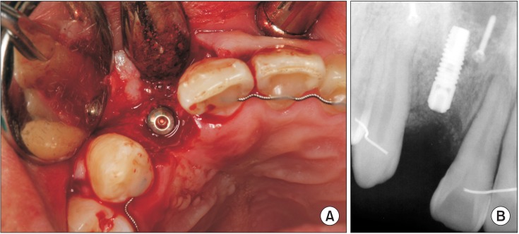
Fig. 3 A. Implant was placed at the expanded ridge. B. Periapical radiograph after implant placement.
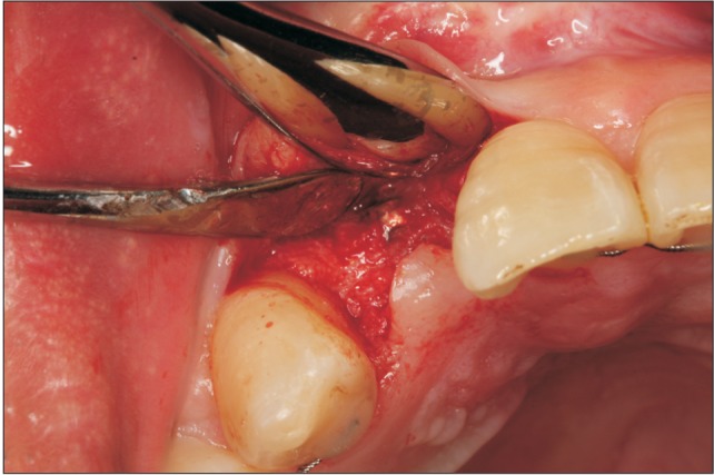
Fig. 4 Clinical view of 2nd surgery.
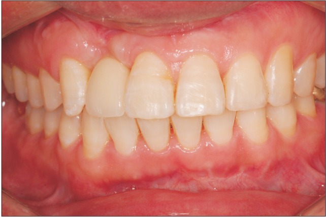
Fig. 5 Delivery of the final implant-supported all-ceramic restoration.
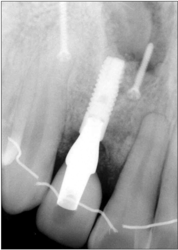
Fig. 6 Periapical view 24 months after the placement of implant.
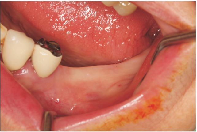
Fig. 7 Clinical aspect before implant placement.
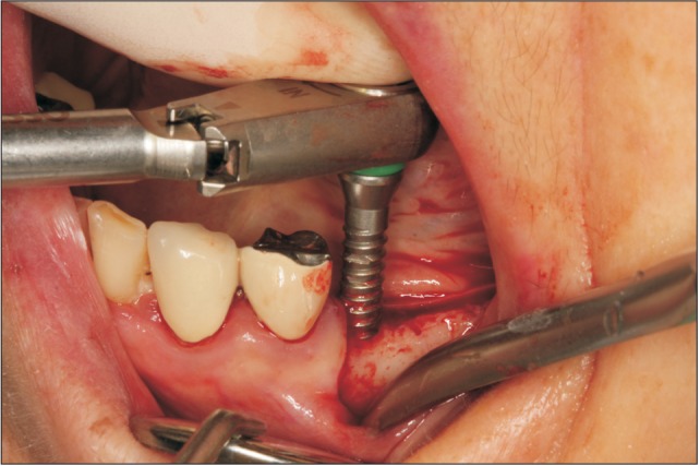
Fig. 8 Expansion of buccal plate achieved by splitting the ridge using the bone expander in SplitMaster (Mr. Curette).
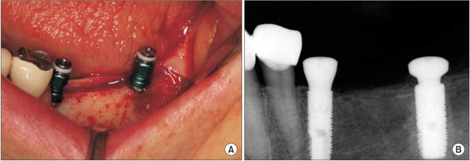
Fig. 9 A. Implants were placed at the expanded ridge. B. Periapical radiograph after implant placement.
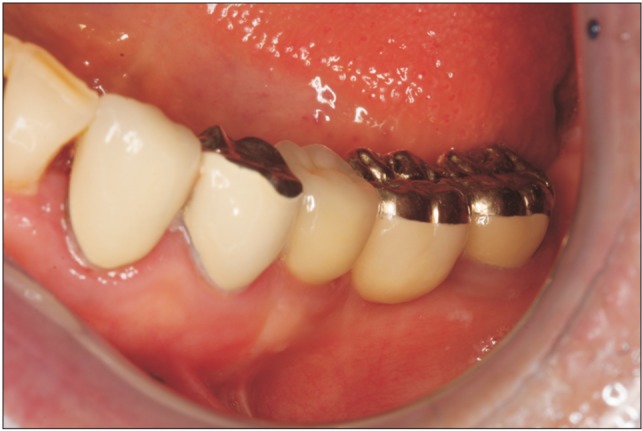
Fig. 10 Intraoral view after delivery of final implant-supported porcelain fused to metal restoration.
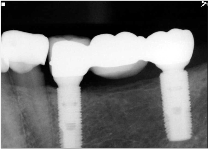
Fig. 11 Periapical radiograph 63 months after implant placement.
Reference
-
1. Buser D, Brägger U, Lang NP, Nyman S. Regeneration and enlargement of jaw bone using guided tissue regeneration. Clin Oral Implants Res. 1990; 1:22–32. PMID: 2099209.
Article2. Jovanovic SA, Spiekermann H, Richter EJ. Bone regeneration around titanium dental implants in dehisced defect sites: a clinical study. Int J Oral Maxillofac Implants. 1992; 7:233–245. PMID: 1398841.3. Misch CM. Maxillofacial donor sites for sinus floor and alveolar reconstruction. In : Jensen OT, editor. The sinus bone graft. 2nd ed. Chicago: Quintessence Publishing;2006. p. 129–143.4. Jensen J, Sindet-Pedersen S. Autogenous mandibular bone grafts and osseointegrated implants for reconstruction of the severely atrophied maxilla: a preliminary report. J Oral Maxillofac Surg. 1991; 49:1277–1287. PMID: 1955919.
Article5. Kim YK, Kim SG, Lee BG. Bone graft and implant. 1st ed. Seoul: Narae Publishing;2007. p. 435–467.6. Jensen OT, Cullum DR, Baer D. Marginal bone stability using 3 different flap approaches for alveolar split expansion for dental implants: a 1-year clinical study. J Oral Maxillofac Surg. 2009; 67:1921–1930. PMID: 19686930.7. Jensen OT, Cullum DR, Baer D. Marginal bone stability using 3 different flap approaches for alveolar split expansion for dental implants: a 1-year clinical study. J Oral Maxillofac Surg. 2009; 67:1921–1930. PMID: 19686930.8. Chiapasco M, Ferrini F, Casentini P, Accardi S, Zaniboni M. Dental implants placed in expanded narrow edentulous ridges with the Extension Crest device. A 1-3-year multicenter follow-up study. Clin Oral Implants Res. 2006; 17:265–272. PMID: 16672021.9. Nishioka RS, Kojima AN. Screw spreading: technical considerations and case report. Int J Periodontics Restorative Dent. 2011; 31:141–147. PMID: 21491013.10. Chan HL, Fu JH, Koticha T, Benavides E, Wang HL. Ridge width gain with screw spreaders: a cadaver study. Implant Dent. 2013; 22:552–558. PMID: 24013399.11. Sethi A, Kaus T. Maxillary ridge expansion with simultaneous implant placement: 5-year results of an ongoing clinical study. Int J Oral Maxillofac Implants. 2000; 15:491–499. PMID: 10960981.12. Ferrigno N, Laureti M. Surgical advantages with ITI TE implants placement in conjunction with split crest technique. 18-month results of an ongoing prospective study. Clin Oral Implants Res. 2005; 16:147–155. PMID: 15777323.
- Full Text Links
-
- Actions
-
Cited
- CITED
-
- Close
- Share
-
- Similar articles
-
- Implant Placement Using Alveolar Ridge Split in Atrophic Maxillary Alveolar Bone
- Ridge split for implant placement in very thin alveolar ridge
- Ridge Augmentation Using Vascularized Interpositional Periosteal- Connective Tissue (VIP-CT) in Conjunction with Anterior Implant Placement in Maxilla: Report of Three Cases
- Horizontal ridge augmentation with porcine bone-derived grafting material: a long-term retrospective clinical study with more than 5 years of follow-up
- Alveolar Ridge Preservation in the Severely Damaged Sockets of the Anterior Maxilla Followed by Delayed Implant Placement
