J Korean Soc Radiol.
2011 Oct;65(4):381-387. 10.3348/jksr.2011.65.4.381.
Tumor Size Evaluation according to the T Component of the Seventh Edition of the International Association for the Study of Lung Cancer's TNM Classification: Interobserver Agreement between Radiologists and Computer-Aided Diagnosis System in Patients with Lung Cancer
- Affiliations
-
- 1Department of Radiology, Chung-Ang University College of Medicine, Seoul, Korea. j3mn@chol.com
- 2Department of Radiology, Kangwon National University School of Medicine, Chuncheon, Korea.
- KMID: 2002939
- DOI: http://doi.org/10.3348/jksr.2011.65.4.381
Abstract
- PURPOSE
To assess the interobserver agreement for tumor size evaluation between radiologists and the computer-aided diagnosis (CAD) system based on the 7th edition of the TNM classification by the International Association for the Study of Lung Cancer in patients with lung cancer.
MATERIALS AND METHODS
We evaluated 20 patients who underwent a lobectomy or pneumonectomy for primary lung cancer. The maximum diameter of each primary tumor was measured by two radiologists and a CAD system on CT, and was staged based on the 7th edition of the TNM classification. The CT size and T-staging of the primary tumors was compared with the pathologic size and staging and the variability in the sizes and T stages of primary tumors was statistically analyzed between each radiologist's measurement or CAD estimation and the pathologic results.
RESULTS
There was no statistically significant interobserver difference for the CT size among the two radiologists, between pathologic and CT size estimated by the radiologists, and between pathologic and CT staging by the radiologists and CAD system. However, there was a statistically significant interobserver difference between pathologic size and the CT size estimated by the CAD system (p = 0.003).
CONCLUSION
No significant differences were found in the measurement of tumor size among radiologists or in the assessment of T-staging by radiologists and the CAD system.
MeSH Terms
Figure
-
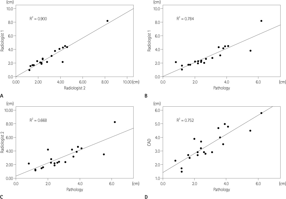
Fig. 1 Comparison of tumor sizes between two radiologists (A), radiologist 1 and pathology (B), radiologist 2 and pathology (C), and CAD and pathology (D). Note.-CAD = computer-aided diagnosis
Reference
-
1. Tsim S, O'Dowd CA, Milroy R, Davidson S. Staging of non-small cell lung cancer (NSCLC): a review. Respir Med. 2010; 104:1767–1774.2. Wang Y, van Klaveren RJ, van der Zaag-Loonen HJ, de Bock GH, Gietema HA, Xu DM, et al. Effect of nodule characteristics on variability of semiautomated volume measurements in pulmonary nodules detected in a lung cancer screening program. Radiology. 2008; 248:625–631.3. Nishino M, Guo M, Jackman DM, DiPiro PJ, Yap JT, Ho TK, et al. CT tumor volume measurement in advanced non-small-cell lung cancer: performance characteristics of an emerging clinical tool. Acad Radiol. 2011; 18:54–62.4. Iwano S, Okada T, Koike W, Matsuo K, Toya R, Yamazaki M, et al. Semi-automatic volumetric measurement of lung cancer using multi-detector CT effects of nodule characteristics. Acad Radiol. 2009; 16:1179–1186.5. Marten K, Auer F, Schmidt S, Kohl G, Rummeny EJ, Engelke C. Inadequacy of manual measurements compared to automated CT volumetry in assessment of treatment response of pulmonary metastases using RECIST criteria. Eur Radiol. 2006; 16:781–790.6. Rusch VW, Asamura H, Watanabe H, Giroux DJ, Rami-Porta R, Goldstraw P. Members of IASLC Staging Committee. The IASLC lung cancer staging project: a proposal for a new international lymph node map in the forthcoming seventh edition of the TNM classification for lung cancer. J Thorac Oncol. 2009; 4:568–577.7. Lee HY, Lee KS, Hwang HS, Lee JW, Ahn MJ, Park K, et al. Molecularly targeted therapy using bevacizumab for non-small cell lung cancer: a pilot study for the new CT response criteria. Korean J Radiol. 2010; 11:618–626.8. Tanoue LT, Detterbeck FC. New TNM classification for non-small-cell lung cancer. Expert Rev Anticancer Ther. 2009; 9:413–423.9. Macpherson RE, Higgins GS, Murchison JT, Wallace WA, Price A, Gaffney S, et al. Non-small-cell lung cancer dimensions: CT-pathological correlation and interobserver variation. Br J Radiol. 2009; 82:421–425.10. Wormanns D, Diederich S, Lentschig MG, Winter F, Heindel W. Spiral CT of pulmonary nodules: interobserver variation in assessment of lesion size. Eur Radiol. 2000; 10:710–713.11. Zhao B, James LP, Moskowitz CS, Guo P, Ginsberg MS, Lefkowitz RA, et al. Evaluating variability in tumor measurements from same-day repeat CT scans of patients with non-small cell lung cancer. Radiology. 2009; 252:263–272.
- Full Text Links
-
- Actions
-
Cited
- CITED
-
- Close
- Share
-
- Similar articles
-
- The International Association for the Study of Lung Cancer Lymph Node Map: A Radiologic Atlas and Review
- Prognostic Factors in Stage IIB Non-Small Cell Lung Cancer according to the 8th Edition of TNM Staging System
- Impact of the New International Association for the Study of Lung Cancer Staging System in Non-Small Cell Lung Cancer: With Comparison to the Union for International Cancer Control 6th Tumor, Node, Metastasis Edition
- Computer-aided Diagnosis for Lung Cancer
- Evaluation of the 7th UICC TNM Staging System of Gastric Cancer
