Calcifying Fibrous Tumor of the Pleura: A Rare Case with an Unusual Presentation on CT and MRI
- Affiliations
-
- 1Department of Radiology and the Research Institute of Radiological Science, Gangnam Severance Hospital, Yonsei University College of Medicine, Seoul, Korea. thkim1@yuhs.ac
- 2Department of Thoracic and Cardiovascular Surgery, Gangnam Severance Hospital, Yonsei University College of Medicine, Seoul, Korea.
- 3Department of Pathology, Gangnam Severance Hospital, Yonsei University College of Medicine, Seoul, Korea.
- KMID: 2002808
- DOI: http://doi.org/10.3348/jksr.2015.72.2.123
Abstract
- Calcifying fibrous tumors (CFTs) are rare benign mesenchymal tumors consisting of hyalinized collagenous fibrotic tissue with a lymphoplasmacytic infiltrate and dystrophic calcifications. Radiographic features have seldom been described, and there are no reports describing magnetic resonance imaging (MRI) findings. Here, we report a pleural CFT in a 47-year-old woman. The tumor mimicked an intrapulmonary lesion on initial computed tomography scans but migrated inferiorly and presented as an extra-pulmonary lesion on MRI. The tumor showed iso-signal intensity on T1-weighted images (WIs), low signal intensity on T2WIs, and slight rim enhancement on enhanced T1WIs.
Figure
-
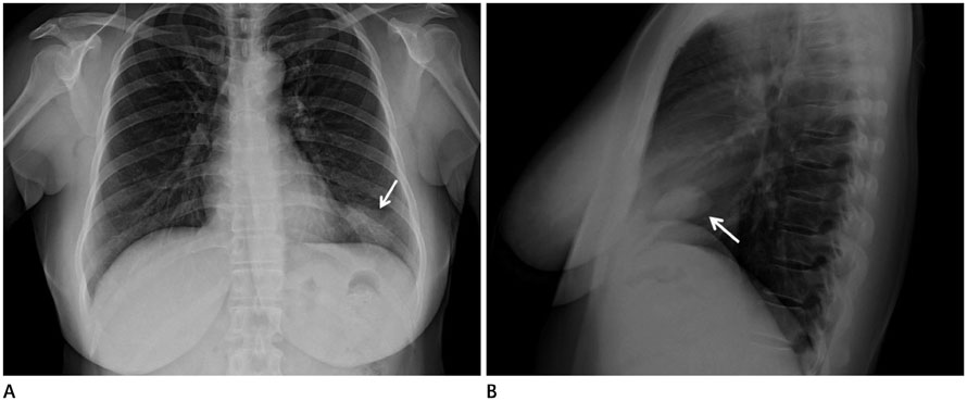
Fig. 1 Plain chest X-ray of the calcifying fibrous tumor of the pleura. Chest PA (A) and lateral view (B) show a 3 × 4.5 cm, oval-shaped, well-circumscribed mass-like lesion (arrow) in the left lower lung periphery.
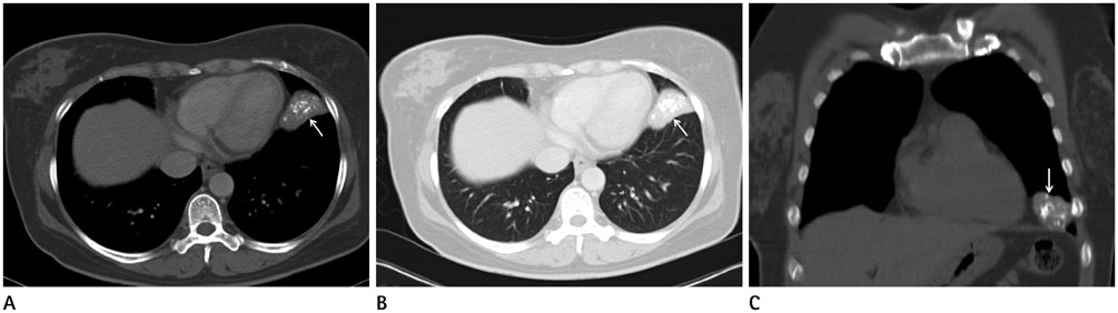
Fig. 2 High-resolution CT image of the calcifying fibrous tumor of the pleura. Axial CT scan on mediastinal (A), lung window setting (B), and coronal view mediastinal setting (C) show a 2.5 × 4.5 cm well-circumscribed, lobulated mass (arrow) with coarse calcification, located above the diaphragm in the lingular segment of the left upper lobe.
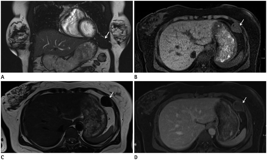
Fig. 3 MR images of the calcifying fibrous tumor of the pleura. The ultrafast spine echo Half-Fourier-acquisition single-shot turbo spine-echo coronal image (A) shows an inferiorly migrated mass located in the extraparenchymal space between the left diaphragm and left lung. The mass (arrow) shows iso- to intermediate signal intensity on the T1-weighted image (B) and low signal intensity on the T2-weighted image (C). Slight peripheral rim enhancement was observed on the gadolinium-enhanced T1-weighted image (D).

Fig. 4 Gross images and histology of the specimen. The mass measured up to 6 cm, well demarcated, firm and lobulated (A). Cut surfaces of the mass revealed a solid, white, fibrous matrix without hemorrhage or necrosis (B). On microscopic exam, fibrotic tissue with dystrophic calcification was observed [hematoxylin and eosin stain (× 100)] (C) with vimentin positive on immunohistochemical staining (× 200) (D).
Reference
-
1. Pinkard NB, Wilson RW, Lawless N, Dodd LG, McAdams HP, Koss MN, et al. Calcifying fibrous pseudotumor of pleura. A report of three cases of a newly described entity involving the pleura. Am J Clin Pathol. 1996; 105:189–119.2. Fetsch JF, Montgomery EA, Meis JM. Calcifying fibrous pseudotumor. Am J Surg Pathol. 1993; 17:502–508.3. Mito K, Kashima K, Daa T, Kondoh Y, Miura T, Kawahara K, et al. Multiple calcifying fibrous tumors of the pleura. Virchows Arch. 2005; 446:78–81.4. Jiang K, Nie J, Wang J, Li J. Multiple calcifying fibrous pseudotumor of the bilateral pleura. Jpn J Clin Oncol. 2011; 41:130–133.5. Isaka M, Nakagawa K, Maniwa T, Saisho S, Ohde Y, Okumura T, et al. Disseminated calcifying tumor of the pleura: review of the literature and a case report with immunohistochemical study of its histogenesis. Gen Thorac Cardiovasc Surg. 2011; 59:579–582.6. Ammar A, El Hammami S, Horchani H, Sellami N, Kilani T. Calcifying fibrous pseudotumor of the pleura: a rare location. Ann Thorac Surg. 2003; 76:2081–2082.7. Jang KS, Oh YH, Han HX, Chon SH, Chung WS, Park CK, et al. Calcifying fibrous pseudotumor of the pleura. Ann Thorac Surg. 2004; 78:e87–e88.8. Ferretti GR, Chiles C, Choplin RH, Coulomb M. Localized benign fibrous tumors of the pleura. AJR Am J Roentgenol. 1997; 169:683–686.9. Shibata K, Yuki D, Sakata K. Multiple calcifying fibrous pseudotumors disseminated in the pleura. Ann Thorac Surg. 2008; 85:e3–e5.
- Full Text Links
-
- Actions
-
Cited
- CITED
-
- Close
- Share
-
- Similar articles
-
- Multiple Calcifying Fibrous Pseudotumors in the Pleura : A case report
- Calcifying fibrous pseudotumor of mediastinum--a case report
- Spontaneous Interlobar Pneumothorax in a Localized Fibrous Tumor of in the Pleura
- Solitary Fibrous Tumor of the Adrenal Gland: A Case Report
- MRI Findings of a Malignant Solitary Fibrous Tumor of the Diaphragmatic Pleura: a Case Report
