Korean J Urol.
2006 Apr;47(4):440-442. 10.4111/kju.2006.47.4.440.
A Fibrotic Nodule in the Corpus Cavernosum
- Affiliations
-
- 1Department of Urology, College of Medicine, The Catholic University of Korea, Seoul, Korea.
- KMID: 1997157
- DOI: http://doi.org/10.4111/kju.2006.47.4.440
Abstract
- Fibrotic lesions occurring in the corpus cavernosum are usually cases of Peyronie's disease that originate from the tunica albuginea, or they are the fibrotic result of inflammatory processes. The lesion involving the corpus cavernosum, but not tunica albuginea is rare. We present here a case of fibrotic nodule arising in the corpus cavernosum with the sonographic and magnetic resonance imaging features. A 38-year-old man complained a small nodular mass in the left corpus cavernosum at the level of penoscrotal junction without abnormal curvature of the organ. We performed ultrasonography and magnetic resonance imaging to determine exactly what the lesion was. The lesion was removed and it was pathologically found to be a localized fibrotic nodule of the corpus cavernosum with some narrow-channeled vascular structures.
Figure
-

Fig. 1 (A) Ultrasonography shows a heterogenous echogenic mass. (B) On MRI, a T2 and T1 low intensity signal nodule is noted on the left corpus cavernosum. This lesion is not definitely enhanced on the post-contrast film.
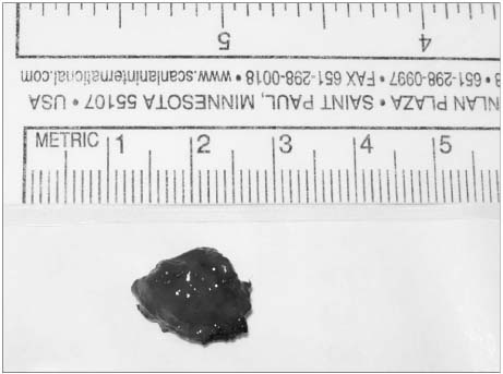
Fig. 2 The totally excised mass from the corpus cavernosum. It is round, reddish and rubbery.
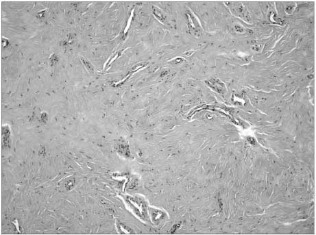
Fig. 3 The microscopic view shows the diffuse fibrosis with some narrow channeled vascular structures.
Reference
-
1. Lopes M, Lanzafame S, Magro G. Localized fibrosis of the corpus cavernosum: an example of fibrosis arising from the vascular smooth muscle cells. Report of a case with histogenetic considerations. Urol Int. 2000. 64:173–177.2. Smith BH. Peyronie's disease. Am J Clin Pathol. 1966. 45:670–678.3. Enzinger FM, Weiss S. Soft tissue tumors. 2001. 4th ed. St Louis: Mosby;45–102.4. Bernardino ME, Jing BS, Thomas JL, Lindell MM Jr, Zornoza J. The extremity soft tissue lesion: a comparative study of ultrasound, computed tomography, and xeroradiography. Radiology. 1981. 139:53–59.5. Hughes TM, Spillane AJ. Imaging of soft tissue tumours. Br J Surg. 2000. 87:259–260.6. Crim JR, Seegar LL, Yao L, Chandnani V, Eckardt JJ. Diagnosis of soft tissue masses with MR imaging: can benign masses be differentiated from malignant ones? Radiology. 1992. 185:581–586.7. Egund N, Ekelund L, Sako M, Persson B. CT of soft-tissue tumors. Am J Roentgenol. 1981. 137:725–729.8. Schurch W, Seemayer T, Gabbiani G, Sternberg SS. Histology for pathologists. 1992. New York: Raven Press;118–125.9. Gabbiani G. The cellular derivation and the life span of the myofibroblast. Pathol Res Pract. 1996. 192:708–711.10. Sappino AP, Schurch W, Gabbiani G. Differentiation repertoire of fibroblastic cells: expression of cytoskeletal proteins as marker of phenotypic modulations. Lab Invest. 1990. 63:144–161.
- Full Text Links
-
- Actions
-
Cited
- CITED
-
- Close
- Share
-
- Similar articles
-
- Pharmacologic Effect of Phentolamine on Norepinephrine Induced Contraction of Corpus Cavernosum
- A Case or Priapism Treated by Corpus Cavernosum-Saphenous Vein Anastomosis
- A Case of Penile Injury (Accidental Rupture of Corpus Cavernosum)
- The Effect and Mechanism of the Specific Phosphodiesterase (PDE) III-Inhibitor Milrinone on Human and Rabbit Corpus Cavernosum
- Priapism: Treatment by corpus cavernosum-saphenous vein anastomosis
