J Korean Acad Conserv Dent.
2007 Jan;32(1):19-27. 10.5395/JKACD.2007.32.1.019.
Proposal of new dental color-space for aesthetic dental materials
- Affiliations
-
- 1Department of Conservative Dentistry, School of Dentistry, DSRI, Chonnam National University, Korea. hinso@jnu.ac.kr
- KMID: 1986927
- DOI: http://doi.org/10.5395/JKACD.2007.32.1.019
Abstract
- The purpose of this study is to develope new dental color-space system. Twelve kinds of dental composites and one kind of dental porcelain were used in this study. Disk samples (15 mm in diameter, 4 mm in thickness) of used materials were made and sample's CIE L*a*b* value was measured by Spectrocolorimeter (MiniScan XE plus, Model 4000S, diffuse/8degrees viewing mode, 14.3 mm Port diameters, Hunter Lab. USA). The range of measured color distribution was analyzed. All the data were applied in the form of T### which is expression unit in CNU Cons Dental Color Chart. The value of L* lies between 80.40 and 52.70. The value of a* are between 10.60 and 3.60 and b* are between 28.40 and 2.21. The average value of L* is 67.40, and median value is 67.30. The value of a* are 2.89 and 2.91 respectively. And for the b*, 14.30 and 13.90 were obtained. The data were converted to T### that is the unit count system in CNU-Cons Dental Color Chart. The value of L* is converted in the first digit of the numbering system. Each unit is 2.0 measured values. The second digit is the value of a* and is converted new number by 1.0 measured value. For the third digit b* is replaced and it is 2.0 measured unit apart. T555 was set to the value of L* ranging from 66.0 to 68.0, value of a* ranging from 3 to 4 and b* value ranging from 14 to 16.
Keyword
Figure
-
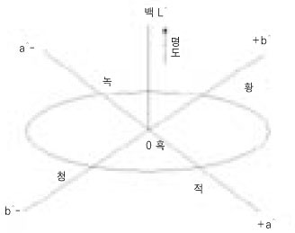
Figure 1 Schematic diagram of the CIE L*a*b* color system.
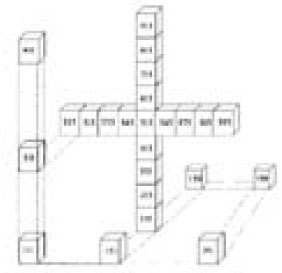
Figure 2 Diagram of the CNU-Cons Dental Color system.

Figure 3 L* and C* value of the tested materials.

Figure 4 a* and b* value of the tested materials.
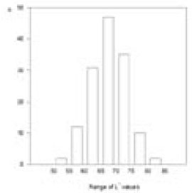
Figure 5 Histogram of the L* value.

Figure 6 Histogram of the a* value.

Figure 7 Histogram of the b* value.
Reference
-
1. Clark EB. An analysis of tooth color. J Am Dent Assoc. 1931. 18:2093–2103.
Article2. Clark EB. Tooth color selection. J Am Dent Assoc. 1933. 20:1065–1073.3. Miller LL. Shade matching. J Esthet Dent. 1993. 5:143–153.
Article4. Miller LL. Organizing color in dentistry. J Am Dent Assoc. 1987. 12. Spec No:26E–40E.
Article5. Hayashi T. Medical color standard. V. Tooth crown. 1967. Tokyo: Japan Color Research Institute.6. Sproull RC. Color matching in dentistry. Part II: Practical applications of the organization of color. J Prosthet Dent. 1973. 29:556–566.
Article7. Lemire PA, Burk B. Color in dentistry. 1975. Hartford: CT J.M Ney Co..8. Grajower R, Revah A, Sorin S. Reflectance spectra of natural and acrylic teeth. J Prosthet Dent. 1976. 36:570–579.9. Macentee M, Lakowski R. Instrumental color measurement of vital and extracted teeth. J Oral Rehabil. 1981. 8:203–208.
Article10. Goodkind RJ, Schwabacher WB. Use of a fiber-optic colorimeter for in vivo color measurements of 2830 anterior teeth. J Prosthet Dent. 1987. 58:535–542.
Article11. Park HG, Jeong JH. A Study on the Color of Korean Natural Teeth. J Korean Acad Prosthodont. 1988. 26:185–196.12. Hwang IN, Oh WM. Colorimetric analysis of extracted human teeth and five shade guides. J Korean Acad Conserv Dent. 1997. 22:769–781.13. Cho GM, Shin DH. Color analysis of the natural teeth with a modified intraoral spectrophotometer. J Korean Acad Conserv Dent. 1998. 23:223–235.14. Hwang IN, Lee GW. Translucency of light cured composite resins depends on thickess & its influence on color of restorations. J Korean Acad Conserv Dent. 1999. 24:604–613.15. Goodkind RJ, Loupe MJ. Teaching of color in predoctoral and postdoctoral dental education in 1988. J Prosthet Dent. 1992. 67:713–717.
Article16. Kim HS, Um JM. A study on color differences between composite resins and shade guides. J Korean Acad Conserv Dent. 1996. 21:107–120.17. Cho GI, Hwang IN, Choi HR, O WM. Comparative evaluation of light-cred composite resins based on vita shade by spectrocolorimeter. J Korean Acad Conserv Dent. 1998. 23:424–432.18. Gross MD, Moser JB. A colorimetric study of coffee and tee staining of four composite resins. J Oral Rehabil. 1977. 4:311–322.
Article19. Seghi RR, Hewlett ER, Kim J. Visual and instrumental colorimetric asessments of small color differences on translucent dental porcelain. J Dent Res. 1989. 68:1760–1764.
Article20. Wozniak WT. Proposed guidelines for the acceptance program for dental shade guides. 1987. Chicago: American Dental Association.21. O'Neal SJ, Powell WD. Color discrimination and shade matching ability of third year dental student. J Prosthet Dent. 1984. 63:174.
- Full Text Links
-
- Actions
-
Cited
- CITED
-
- Close
- Share
-
- Similar articles
-
- Digital shade and camera use in dental practice
- Spectrophotometic analysis of the influence of substrate on the color of dental ceramics
- The effect of repeated firings on the color change and surface roughness of dental ceramics
- Comparison of the Surface Properties and Color Stability of Various Aesthetic Restorative Materials Treated with In-Office Tooth Bleaching
- A comparative study on color and dimensional stability of esthetic indirect dental materials
