Resorption of labial bone in maxillary anterior implant
- Affiliations
-
- 1Department of Prosthodontics, Graduate School of Chosun University, Gwangju, Korea. khjdds@chosun.ac.kr
- KMID: 1975082
- DOI: http://doi.org/10.4047/jap.2011.3.2.85
Abstract
- PURPOSE
The purpose of this study was to evaluate the amount of resorption and thickness of labial bone in anterior maxillary implant using cone beam computed tomography with Hitachi CB Mercuray (Hitachi, Medico, Tokyo, Japan).
MATERIALS AND METHODS
Twenty-one patients with 26 implants were followed-up and checked with CBCT. 21 OSSEOTITE NT(R) (3i/implant Innovations, Florida, USA) and 5 OSSEOTITE(R) implants (3i/implant Innovations, Florida, USA) were placed at anterior region and they were positioned vertically at the same level of bony scallop of adjacent teeth. Whenever there was no lesion or labial bone was intact, immediate placement was tried as possible as it could be. Generated bone regeneration was done in the patients with the deficiency of hard tissue using Bio-Oss(R) (Geistlich, Wolhusen, Switzerland) and Bio-Gide(R) (Geistlich, Wolhusen, Switzerland). Second surgery was done in 6 months after implant placement and provisionalization was done for 3 months. Definite abutment was made of titanium abutment with porcelain, gold and zirconia, and was attached after provisionalization. Two-dimensional slices were created to produce sagittal, coronal, axial and 3D by using OnDemand3D (Cybermed, Seoul, Korea).
RESULTS
The mean value of bone resorption (distance from top of implant to labial bone) was 1.32 +/- 0.86 mm and the mean thickness of labial bone was 1.91 +/- 0.45 mm.
CONCLUSION
It is suggested that the thickness more than 1.91 mm could reduce the amount and incidence of resorption of labial bone in maxillary anterior implant.
MeSH Terms
Figure
-
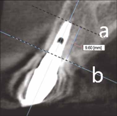
Fig. 1 Determination of the position of platform (Baseline). a: Apex of implant, b: platform of implant, a - b: length of implant.

Fig. 2 Measurement of the amount of labial bone loss. a: apex of implant, b: platform of implant, c: position of labial bone, L1: length of implant, L2: length from apex to labial bone, Ra (= L1 - L2): the amount of labial bone loss.
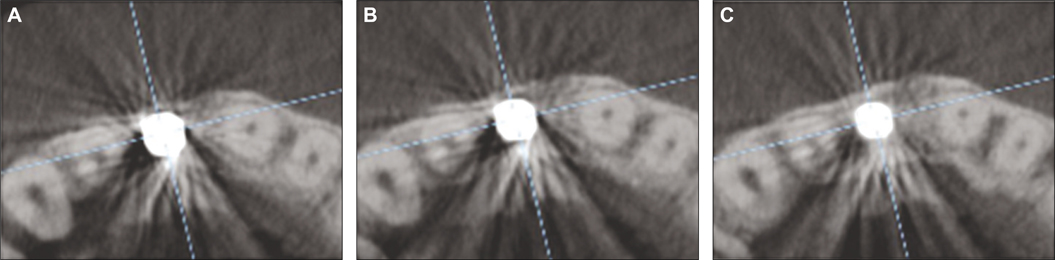
Fig. 3 Determination of the existence of labial bone. A: absence of labial bone, B: labial bone seemed to exist but indistinct, C: labial bone and continuity of labial bone with adjacent tooth were clear (distinct).
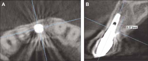
Fig. 4 Determination of the position of labial bone. A: clear labial bone and its continuity, B: the position of labial bone at (A), L2 minus L1 means the amount of labial bone loss in CBCT.

Fig. 5 Measurement of the thickness of labial bone.
Cited by 3 articles
-
The thickness of alveolar bone at the maxillary canine and premolar teeth in normal occlusion
Seong-Ho Jin, Jun-Beom Park, Namryang Kim, Seojin Park, Kyung Jae Kim, Yoonji Kim, Yoon-Ah Kook, Youngkyung Ko
J Periodontal Implant Sci. 2012;42(5):173-178. doi: 10.5051/jpis.2012.42.5.173.Influence of the anterior arch shape and root position on root angulation in the maxillary esthetic area
Suweera Petaibunlue, Pravej Serichetaphongse, Atiphan Pimkhaokham
Imaging Sci Dent. 2019;49(2):123-130. doi: 10.5624/isd.2019.49.2.123.Radiographic evaluation of computer aided design/computer aided manufacturing (CAD/CAM) customized abutment of implant
Tae-Gyeong Yun, Gyeong-Je Lee, Chae-Heon Chung, Hee-Jung Kim
J Korean Acad Prosthodont. 2017;55(3):258-263. doi: 10.4047/jkap.2017.55.3.258.
Reference
-
1. Garber DA. The esthetic dental implant: letting restoration be the guide. J Oral Implantol. 1996. 22:45–50.2. Tarnow DP, Eskow RN, Zamzok J. Aesthetics and implant dentistry. Periodontol 2000. 1996. 11:85–94.3. Becker W, Becker BE. Guided tissue regeneration for implants placed into extraction sockets and for implant dehiscences: surgical techniques and case report. Int J Periodontics Restorative Dent. 1990. 10:376–391.4. Bahat O, Fontanesi RV, Preston J. Reconstruction of the hard and soft tissues for optimal placement of osseointegrated implants. Int J Periodontics Restorative Dent. 1993. 13:255–275.5. Kan JY, Rungcharassaeng K. Site development for anterior single implant esthetics: the dentulous site. Compend Contin Educ Dent. 2001. 22:221–226. 228230–231.6. Wöhrle PS. Nobel Perfect esthetic scalloped implant: rationale for a new design. Clin Implant Dent Relat Res. 2003. 5:64–73.7. Lazzara RJ, Porter SS. Platform switching: a new concept in implant dentistry for controlling postrestorative crestal bone levels. Int J Periodontics Restorative Dent. 2006. 26:9–17.8. Schrotenboer J, Tsao YP, Kinariwala V, Wang HL. Effect of microthreads and platform switching on crestal bone stress levels: a finite element analysis. J Periodontol. 2008. 79:2166–2172.9. Lindquist LW, Carlsson GE, Jemt T. A prospective 15-year follow-up study of mandibular fixed prostheses supported by osseointegrated implants. Clinical results and marginal bone loss. Clin Oral Implants Res. 1996. 7:329–336.10. Jung YC, Han CH, Lee KW. A 1-year radiographic evaluation of marginal bone around dental implants. Int J Oral Maxillofac Implants. 1996. 11:811–818.11. Small PN, Tarnow DP. Gingival recession around implants: a 1-year longitudinal prospective study. Int J Oral Maxillofac Implants. 2000. 15:527–532.12. Bengazi F, Wennström JL, Lekholm U. Recession of the soft tissue margin at oral implants. A 2-year longitudinal prospective study. Clin Oral Implants Res. 1996. 7:303–310.13. Cecchinato D, Bengazi F, Blasi G, Botticelli D, Cardarelli I, Gualini F. Bone level alterations at implants placed in the posterior segments of the dentition: outcome of submerged/non-submerged healing. A 5-year multicenter, randomized, controlled clinical trial. Clin Oral Implants Res. 2008. 19:429–431.14. Albrektsson T, Zarb G, Worthington P, Eriksson AR. The long-term efficacy of currently used dental implants: a review and proposed criteria of success. Int J Oral Maxillofac Implants. 1986. 1:11–25.15. Oh TJ, Yoon J, Misch CE, Wang HL. The causes of early implant bone loss: myth or science? J Periodontol. 2002. 73:322–333.16. Hermann JS, Schoolfield JD, Nummikoski PV, Buser D, Schenk RK, Cochran DL. Crestal bone changes around titanium implants: a methodologic study comparing linear radiographic with histometric measurements. Int J Oral Maxillofac Implants. 2001. 16:475–485.17. Persson LG, Lekholm U, Leonhardt A, Dahlén G, Lindhe J. Bacterial colonization on internal surfaces of Brånemark system implant components. Clin Oral Implants Res. 1996. 7:90–95.18. Guindy JS, Besimo CE, Besimo R, Schiel H, Meyer J. Bacterial leakage into and from prefabricated screw-retained implant-borne crowns in vitro. J Oral Rehabil. 1998. 25:403–408.19. Broggini N, McManus LM, Hermann JS, Medina RU, Oates TW, Schenk RK, Buser D, Mellonig JT, Cochran DL. Persistent acute inflammation at the implant-abutment interface. J Dent Res. 2003. 82:232–237.20. Cochran DL, Hermann JS, Schenk RK, Higginbottom FL, Buser D. Biologic width around titanium implants. A histometric analysis of the implanto-gingival junction around unloaded and loaded nonsubmerged implants in the canine mandible. J Periodontol. 1997. 68:186–198.21. Hermann JS, Buser D, Schenk RK, Higginbottom FL, Cochran DL. Biologic width around titanium implants. A physiologically formed and stable dimension over time. Clin Oral Implants Res. 2000. 11:1–11.22. Hermann JS, Buser D, Schenk RK, Schoolfield JD, Cochran DL. Biologic Width around one- and two-piece titanium implants. Clin Oral Implants Res. 2001. 12:559–571.23. Abrahamsson I, Berglundh T, Lindhe J. The mucosal barrier following abutment dis/reconnection. An experimental study in dogs. J Clin Periodontol. 1997. 24:568–572.24. Benkow HH. A new principle and appliance for radiographic tooth measurements. J Dent Res. 1957. 36:641–643.25. Rosling B, Hollender L, Nyman S, Olsson G. A radiographic method for assessing changes in alveolar bone height following periodontal therapy. J Clin Periodontol. 1975. 2:211–217.26. Scarfe WC, Farman AG, Sukovic P. Clinical applications of conebeam computed tomography in dental practice. J Can Dent Assoc. 2006. 72:75–80.27. Draenert FG, Coppenrath E, Herzog P, Müller S, Mueller-Lisse UG. Beam hardening artefacts occur in dental implant scans with the NewTom cone beam CT but not with the dental 4-row multidetector CT. Dentomaxillofac Radiol. 2007. 36:198–203.28. Davarpanah M, Martinez H, Etienne D, Zabalegui I, Mattout P, Chiche F, Michel JF. A prospective multicenter evaluation of 1,583 3i implants: 1- to 5-year data. Int J Oral Maxillofac Implants. 2002. 17:820–828.29. Levin L, Pathael S, Dolev E, Schwartz-Arad D. Aesthetic versus surgical success of single dental implants: 1- to 9-year follow-up. Pract Proced Aesthet Dent. 2005. 17:533–538.30. Esposito M, Ekestubbe A, Gröndahl K. Radiological evaluation of marginal bone loss at tooth surfaces facing single Brånemark implants. Clin Oral Implants Res. 1993. 4:151–157.31. Hänggi MP, Hänggi DC, Schoolfield JD, Meyer J, Cochran DL, Hermann JS. Crestal bone changes around titanium implants. Part I: A retrospective radiographic evaluation in humans comparing two non-submerged implant designs with different machined collar lengths. J Periodontol. 2005. 76:791–802.32. Spray JR, Black CG, Morris HF, Ochi S. The influence of bone thickness on facial marginal bone response: stage 1 placement through stage 2 uncovering. Ann Periodontol. 2000. 5:119–128.
- Full Text Links
-
- Actions
-
Cited
- CITED
-
- Close
- Share
-
- Similar articles
-
- Strategy of Esthetic Implant Restoration in Maxillary Anterior Teeth with Severely Resorbed Labial Bone Plate: A Case Report
- Radiographic Study of Peri-Implant Bone Loss and Its Relationship to the Morphology on Maxillary Anterior Alveolar Ridge
- Angulation between Long Axis of Anterior Teeth and Alveolar Process, and Thickness of Alveolar Bone
- Aesthetic implant restoration with alveolar bone graft and digital method on maxillary central incisor: a case report
- Horizontal alteration of anterior alveolar ridge after immediate implant placement: A retrospective cone beam computed tomography analysis
