J Adv Prosthodont.
2014 Aug;6(4):317-324. 10.4047/jap.2014.6.4.317.
Effects of metal surface grinding at the porcelain try-in stage of fixed dental prostheses
- Affiliations
-
- 1Department of Prosthodontics, Faculty of Dentistry, Erciyes University, Kayseri, Turkey. hikilinc@erciyes.edu.tr
- 2Department of Mechatronics Engineering, Faculty of Engineering, Erciyes University, Kayseri, Turkey.
- KMID: 1974852
- DOI: http://doi.org/10.4047/jap.2014.6.4.317
Abstract
- PURPOSE
This study was to evaluate the effect of grinding of the inner metal surface during the porcelain try-in stage on metal-porcelain bonding considering the maximum temperature and the vibration of samples.
MATERIALS AND METHODS
Ninety-one square prism-shaped (1 x 1 x 1.5 mm) nickel-chrome cast frameworks 0.3 mm thick were prepared. Porcelain was applied on two opposite outer axial surfaces of the frameworks. The grinding was performed from the opposite axial sides of the inner metal surfaces with a low-speed handpiece with two types of burs (diamond, tungsten-carbide) under three grinding forces (3.5 N, 7 N, 14 N) and at two durations (5 seconds, 10 seconds). The shear bond strength (SBS) test was performed with universal testing machine. Statistical analyzes were performed at 5% significance level.
RESULTS
The samples subjected to grinding under 3.5 N showed higher SBS values than those exposed to grinding under 7 N and 14 N (P<.05). SBS values of none of the groups differed from those of the control group (P>.05). The types of bur (P=.965) and the duration (P=.679) did not affect the SBS values. On the other hand, type of bur, force applied, and duration of the grinding affected the maximum temperatures of the samples, whereas the maximum vibration was affected only by the type of bur (P<.05).
CONCLUSION
Grinding the inner metal surface did not affect the metal-porcelain bond strength. Although the grinding affected the maximum temperature and the vibration values of the samples, these did not influence the bonding strength.
Figure
-
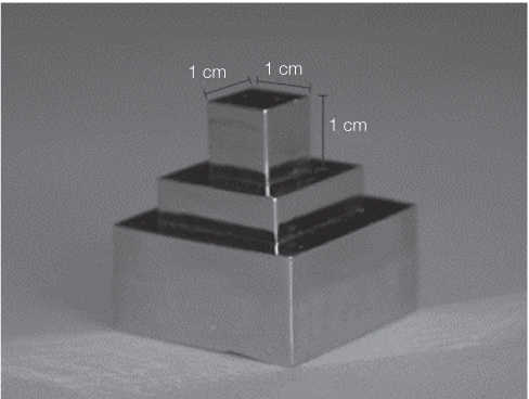
Fig. 1 Template for fabrication of standart metallic frameworks.

Fig. 2 Standardizing unfired porcelain (b) forming by sliding template #2 (a) on metallic framework (c) vertically (A) and horizontally (B).
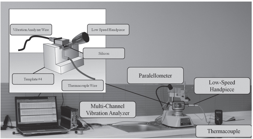
Fig. 3 Experimental setup.

Fig. 4 (A) Preparation of wax pattern on investment duplicate; (B) Finished metal framework.

Fig. 5 Mean and standard deviation shear bond strength graphs for all groups.
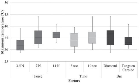
Fig. 6 Maximum temperature box-plot graphs of all groups.
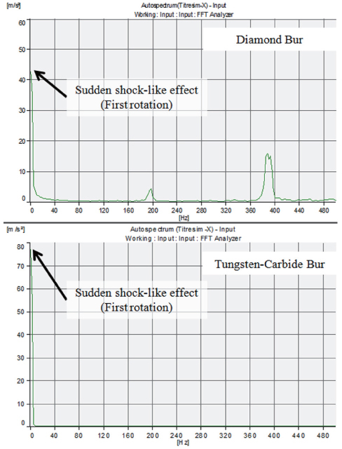
Fig. 7 Grinding vibration graphs for the x axes (Maximum vibration values were pointed out with arrows).

Fig. 8 Maximum vibration box-plot graphs of all groups for the x axes.
Reference
-
1. Lombardo GH, Nishioka RS, Souza RO, Michida SM, Kojima AN, Mesquita AM, Buso L. Influence of surface treatment on the shear bond strength of ceramics fused to cobalt-chromium. J Prosthodont. 2010; 19:103–111.2. Shillingburg HT, Hobo S, Whitsett LD, Jacobi R, Brackett SE. Fundamentals of Fixed Prosthodontics. 3rd ed. Chicago: Quintessence Publishing Co., Inc.;1997. p. 455–484.3. do Prado RA, Panzeri H, Fernandes Neto AJ, das Neves FD, da Silva MR, Mendonça G. Shear bond strength of dental porcelains to nickel-chromium alloys. Braz Dent J. 2005; 16:202–206.4. Shillingburg HT Jr, Hobo S, Fisher DW. Preparation design and margin distortion in porcelain-fused-to-metal restorations. J Prosthet Dent. 1973; 29:276–284.5. Faucher RR, Nicholls JI. Distortion related to margin design in porcelain-fused-to-metal restorations. J Prosthet Dent. 1980; 43:149–155.6. Tao J, Han D. The effect of finish line curvature on marginal fit of all-ceramic CAD/CAM crowns and metal-ceramic crowns. Quintessence Int. 2009; 40:745–752.7. Gemalmaz D, Alkumru HN. Marginal fit changes during porcelain firing cycles. J Prosthet Dent. 1995; 73:49–54.8. Papazoglou E, Brantley WA, Johnston WM. Evaluation of high-temperature distortion of high-palladium metal-ceramic crowns. J Prosthet Dent. 2001; 85:133–140.9. Ushiwata O, de Moraes JV, Bottino MA, da Silva EG. Marginal fit of nickel-chromium copings before and after internal adjustments with duplicated stone dies and disclosing agent. J Prosthet Dent. 2000; 83:634–643.10. Keys LG. An alternate method of verifying the seating of all-ceramic restorations. J Prosthet Dent. 2002; 87:411.11. Song XF, Yin L, Han YG, Wang H. In vitro rapid intraoral adjustment of porcelain prostheses using a high-speed dental handpiece. Acta Biomater. 2008; 4:414–424.12. Kourtis SG, Tripodakis AP, Doukoudakis AA. Spectrophotometric evaluation of the optical influence of different metal alloys and porcelains in the metal-ceramic complex. J Prosthet Dent. 2004; 92:477–485.13. Aladağ A, Cömlekoğlu ME, Dündar M, Güngör MA, Artunç C. Effects of soldering and laser welding on bond strength of ceramic to metal. J Prosthet Dent. 2011; 105:28–34.14. de Vasconcellos LG, Buso L, Lombardo GH, Souza RO, Nogueira L Jr, Bottino MA, Ozcan M. Opaque layer firing temperature and aging effect on the flexural strength of ceramic fused to cobalt-chromium alloy. J Prosthodont. 2010; 19:471–477.15. ISO 9693-1:2012. Dentistry--Compatibility testing-Part 1: Metal-ceramic systems. Geneva: Switerland: International Organization for Standardization;2013.16. Hero H, Syverud M. Carbon impurities and properties of some palladium alloys for ceramic veneering. Dent Mater. 1985; 1:106–110.17. Hammad IA, Talic YF. Designs of bond strength tests for metal-ceramic complexes: review of the literature. J Prosthet Dent. 1996; 75:602–608.18. Brackett SE, Leary JM, Turner KA, Jordan RD. An evaluation of porcelain strength and the effect of surface treatment. J Prosthet Dent. 1989; 61:446–451.19. Leinfelder KF. Porcelain esthetics for the 21st century. J Am Dent Assoc. 2000; 131:47S–51S.20. Isgrò G, Pallav P, van der Zel JM, Feilzer AJ. The influence of the veneering porcelain and different surface treatments on the biaxial flexural strength of a heat-pressed ceramic. J Prosthet Dent. 2003; 90:465–473.
- Full Text Links
-
- Actions
-
Cited
- CITED
-
- Close
- Share
-
- Similar articles
-
- The effect of surface finishes on flexural strength, fracture toughness of feldspathic dental porcelain
- Comparison of retentive force of repair resin by various surface treatment methods in the repair of fractured porcelain fused to metal crown
- Effect of metal conditioner on bonding of porcelain to cobalt-chromium alloy
- Shear bond strength of porcelain repair resins to nonprecious ceramo-metal alloy
- MARGINAL FITNESS OF PORCELAIN-FUSED-TO-METAL CROWN ACCORDING TO MATERIAL AND TECHNIQUE
