Glandular odontogenic cyst: A case report
- Affiliations
-
- 1Department of Oral Medicine and Radiology, Nair Hospital Dental College, Mumbai, India. shezt3@gmail.com
- 2Department of Oral Pathology, Nair Hospital Dental College, Mumbai, India.
- KMID: 1974477
- DOI: http://doi.org/10.5624/isd.2014.44.1.75
Abstract
- Glandular odontogenic cysts (GOCs) are rare intrabony solitary or multiloculated cysts of odontogenic origin. The importance of GOCs lies in the fact that they exhibit a propensity for recurrence similar to keratocystic odontogenic tumors and that they may be confused microscopically with central mucoepidermoid carcinoma. Thus, the oral and maxillofacial radiologists play an important role in definitive diagnosis of GOC based on distinctive cases; though they are rare. In large part, this is due to the GOC's complex and frequently non-specific histopathology. This report describes a case of GOC occurrence in the posterior mandibular ramus region in a 17-year-old female, which is a rare combination of site, age, and gender for occurrence.
MeSH Terms
Figure
-
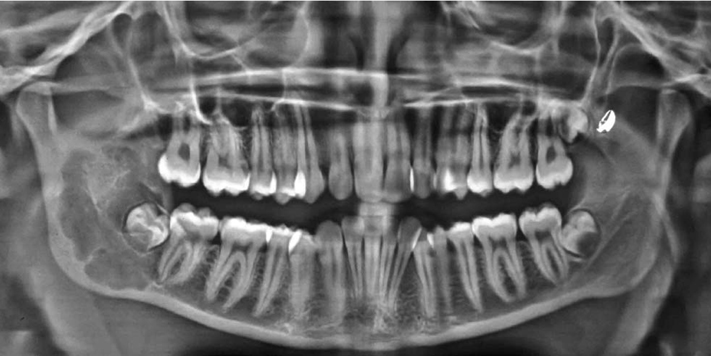
Fig. 1 A panoramic radiograph shows a well-defined multiloculated radiolucent lesion extending from the distal border and apex of lower right second molar into the ramus till approximately 3 mm away from the sigmoid notch enclosing the developing tooth bud of right lower third molar.
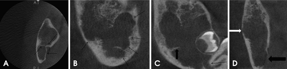
Fig. 2 A. An axial CBCT image shows multiple curved bony septae (black arrows). B. A sagittal CBCT image shows scalloped borders of the cystic lesion (black arrows). C. A reconstructed panoramic CBCT image shows a well-defined radiolucent lesion in the right mandibular ramal region extending from the right lower second molar to the upper third of the ramus with resorption of the apex of the second molar (black arrow) and with well-defined sharp right angle septa (black arrow head). D. A coronal CBCT image shows perforation of the right lingual cortical plate (black arrow head) and thinning of the right buccal cortical plate (white arrow head).
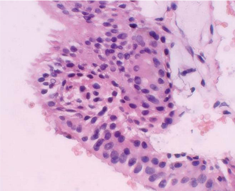
Fig. 3 Histopathologic examination shows cystic lumen lined by pseudo-stratified columnar epithelium with filiform extensions of the cytoplasm and mucous-secreting cells with intra-epithelial spherule formation, characteristic of glandular odontogenic cyst (H&E stain, 400×).
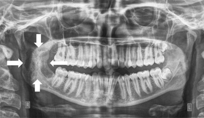
Fig. 4 A panoramic radiograph shows healing of the surgical defect by new bone formation 3 months post-surgery (white arrow heads).
Cited by 1 articles
-
Glandular odontogenic cyst mimicking ameloblastoma in a 78-year-old female: A case report
Byung-Do Lee, Wan Lee, Kyung-Hwan Kwon, Moon-Ki Choi, Eun-Joo Choi, Jung-Hoon Yoon
Imaging Sci Dent. 2014;44(3):249-252. doi: 10.5624/isd.2014.44.3.249.
Reference
-
1. Padayachee A, Van Wyk CW. Two cystic lesions with features of both the botryoid odontogenic cyst and the central mucoepidermoid tumour: sialo-odontogenic cyst? J Oral Pathol. 1987; 16:499–504.
Article2. Gardner DG, Kessler HP, Morency R, Schaffner DL. The glandular odontogenic cyst: an apparent entity. J Oral Pathol. 1988; 17:359–366.
Article3. Kramer IR, Pindborg JJ, Shear M. The WHO histological typing of odontogenic tumours: a commentary on the second edition. Cancer. 1992; 70:2988–2994.4. Sadeghi EM, Weldon LL, Kwon PH, Sampson E. Mucoepidermoid odontogenic cyst. Int J Oral Maxillofac Surg. 1991; 20:142–143.
Article5. High AS, Main DM, Khoo SP, Pedlar J, Hume WJ. The polymorphous odontogenic cyst. J Oral Pathol Med. 1996; 25:25–31.
Article6. Kaplan I, Anavi Y, Hirshberg A. Glandular odontogenic cyst: a challenge in diagnosis and treatment. Oral Dis. 2008; 14:575–581.
Article7. Guruprasad Y, Chauhan DS. Glandular odontogenic cyst of maxilla. J Clin Imaging Sci. 2011; 1:54.
Article8. Nair RG, Varghese IV, Shameena PM, Sudha S. Glandular odontogenic cyst: report of a case and review of literature. J Oral Maxillofac Pathol. 2006; 10:20–23.
Article9. Gardner DG, Morency R. The glandular odontogenic cyst, a rare lesion that tends to recur. J Can Dent Assoc. 1993; 59:929–930.10. Krishnamurthy A, Sherlin HJ, Ramalingam K, Natesan A, Premkumar P, Ramani P, et al. Glandular odontogenic cyst: report of two cases and review of literature. Head Neck Pathol. 2009; 3:153–158.
Article11. Manor R, Anavi Y, Kaplan I, Calderon S. Radiological features of glandular odontogenic cyst. Dentomaxillofac Radiol. 2003; 32:73–79.
Article12. Brannon RB, Kessler HP, Castle JT, Kahn MA. Glandular odontogenic cyst: analysis of 46 cases with special emphasis on microscopic criteria for diagnosis. Head Neck Pathol. 2011; 5:364–375.13. Prabhu S, Rekha K, Kumar G. Glandular odontogenic cyst mimicking central mucoepidermoid carcinoma. J Oral Maxillofac Pathol. 2010; 14:12–15.
Article14. Salehinejad J, Saghafi S, Zare-Mahmoodabadi R, Ghazi N, Kermani H. Glandular odontogenic cyst of the posterior maxilla. Arch Iran Med. 2011; 14:416–418.15. Łuczak K, Nowak R, Rzeszutko M. Glandular odontogenic cyst of the mandible associated with impacted tooth: report of a case and review of literature. Dent Med Probl. 2007; 44:403–406.16. Kaplan I, Anavi Y, Manor R, Sulkes J, Calderon S. The use of molecular markers as an aid in the diagnosis of glandular odontogenic cyst. Oral Oncol. 2005; 41:895–902.
Article17. Noffke C, Raubenheimer EJ. The glandular odontogenic cyst: clinical and radiological features; review of the literature and report of nine cases. Dentomaxillofac Radiol. 2002; 31:333–338.
Article18. MacDonald-Jankowski DS. Glandular odontogenic cyst: systematic review. Dentomaxillofac Radiol. 2010; 39:127–139.
Article
- Full Text Links
-
- Actions
-
Cited
- CITED
-
- Close
- Share
-
- Similar articles
-
- Glandular odontogenic cyst : Report of three cases
- An Unusual Odontogenic Cyst with Diverse Histologic Features
- Glandular odontogenic cyst of mandible: case report
- Clinical and histopathologic analysis of glandular odontogenic cysts of the jaws
- A huge glandular odontogenic cyst occurring at posterior mandible
