Adenomatoid odontogenic tumor of the mandible with unusual radiographic features: A case report
- Affiliations
-
- 1Department of Oral Medicine and Radiology, Coorg Institute of Dental Sciences, Virajpet, India. veesn@rediffmail.com
- 2Department of Oral Medicine and Radiology, Peoples' Dental Academy, Bhopal, India.
- 3Department of Oral Pathology and Microbiology, Peoples' Dental Academy, Bhopal, India.
- KMID: 1974438
- DOI: http://doi.org/10.5624/isd.2013.43.2.111
Abstract
- Adenomatoid odontogenic tumor (AOT) usually presents as a unilocular, pericoronal radiolucency in the maxillary anterior region in adolescent females. Very few conditions occur in such a narrow age range and at such a restrictive site. Rarely, these tumors present with varied clinical features. A case of AOT of the mandible is reported with unusual features such as large size, multilocular appearance, and aggressive behavior. The role of radiology in diagnosis of atypical AOT is extremely important. The unique radiological manifestations of the lesion helped in the diagnosis, and it was managed conservatively with no evidence of recurrence.
MeSH Terms
Figure
-
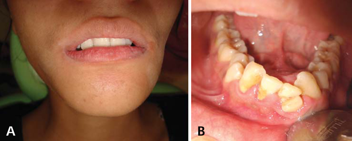
Fig. 1 A. An extraoral photograph shows swelling of the mandible. B. Intraoral photograph shows swelling in the anterior mandible.
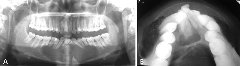
Fig. 2 A. A panoramic radiograph shows the large radiolucent lesion of the mandible with the impacted and displaced left mandibular incisor and root resorption. B. A mandibular cross-sectional occlusal radiograph reveals buccolingual expansion, thinning, and erosion of the cortices, multiple scattered radio-opaque foci, and dense septae.
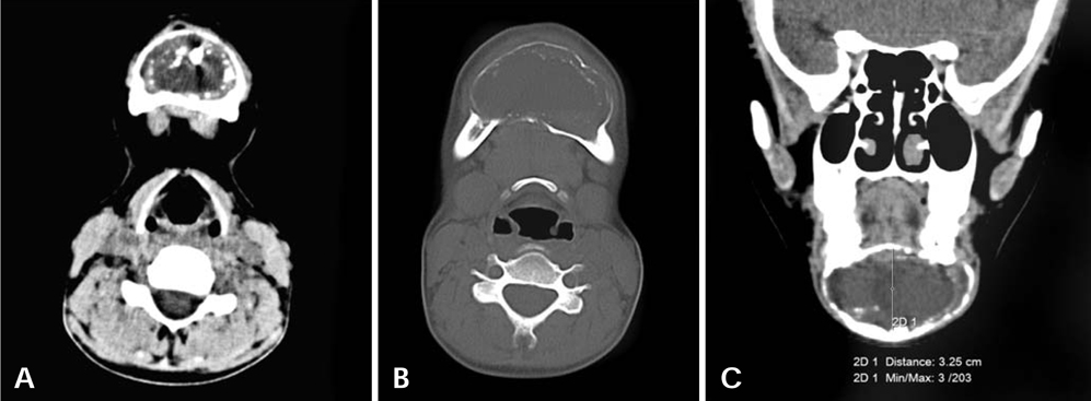
Fig. 3 A. An axial CT image shows the expansile radiolucent lesion with multiple radiopaque flecks towards the periphery. B. An axial CT image of the bone window shows the thinning and discontinuity of the lingual and buccal cortices. C. A coronal CT image shows a large expansile lesion and the soft tissue capsule at the periphery.
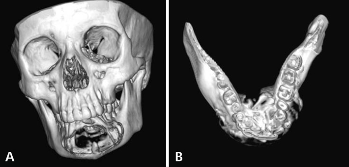
Fig. 4 A. A three-dimensional CT image (anterior view) depicts the lesion in the mandible and the position of the impacted tooth. B. A three-dimensional CT image (occlusal view) reveals the dense lingual septae and multilocular appearance.

Fig. 5 A photomicrograph shows the odontogenic epithelium arranged in whorls and a duct-like arrangement along with the foci of basophilic calcifications (H&E stain, 40×).
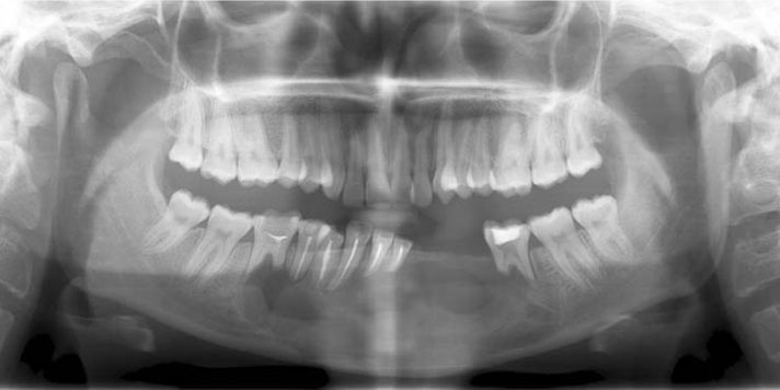
Fig. 6 A six month postoperative panoramic radiograph shows normal healing and no signs of recurrence.
Reference
-
1. White SC, Pharoah MJ. Oral radiology: principles and interpretation. 2004. 5th ed. St. Louis: Mosby.2. Raubenheimer EJ, Seeliger JE, van Heerden WF, Dreyer AF. Adenomatoid odontogenic tumour: a report of two large lesions. Dentomaxillofac Radiol. 1991. 20:43–45.
Article3. Takigami M, Uede T, Imaizumi T, Ohtaki M, Tanabe S, Hashi K. A case of adenomatoid odontogenic tumor with intracranial extension. No Shinkei Geka. 1988. 16:775–779.4. Philipsen HP, Reichart PA. Adenomatoid odontogenic tumour: facts and figures. Oral Oncol. 1999. 35:125–131.
Article5. Dare A, Yamaguchi A, Yoshiki S, Okano T. Limitation of panoramic radiography in diagnosing adenomatoid odontogenic tumors. Oral Surg Oral Med Oral Pathol. 1994. 77:662–668.
Article6. Philipsen HP, Reichart PA, Zhang KH, Nikai H, Yu QX. Adenomatoid odontogenic tumor: biological profile based on 499 cases. J Oral Pathol Med. 1991. 20:149–158.7. Takahashi K, Yoshino T, Hashimoto S. Unusually large cystic adenomatoid odontogenic tumour of the maxilla: case report. Int J Oral Maxillofac Surg. 2001. 30:173–175.
Article8. Mohamed A, Singh AS, Raubenheimer EJ, Bouckaert MM. Adenomatoid odontogenic tumour: review of the literature and an analysis of 33 cases from South Africa. Int J Oral Maxillofac Surg. 2010. 39:843–846.
Article9. Langlais RP, Langland OE, Nortje CJ. Diagnostic imaging of the jaws. 1995. Baltimore: Williams & Wilkins;25.10. Bartake AR, Punnya VA, Sudeendra P, Rekha K. Two adenomatoid odontogenic tumours of the maxilla: a case report. Br J Oral Maxillofac Surg. 2009. 47:638–640.
Article11. Dayi E, Gurbuz G, Bilge OM, Ciftcioglu MA. Adenomatoid odontogenic tumour (adenoameloblastoma). Case report and review of the literature. Aust Dent J. 1997. 42:315–318.
Article12. Nomura M, Tanimoto K, Takata T, Shimosato T. Mandibular adenomatoid odontogenic tumor with unusual clinicopathologic features. J Oral Maxillofac Surg. 1992. 50:282–285.
Article13. Nigam S, Gupta SK, Chaturvedi KU. Adenomatoid odontogenic tumor - a rare cause of jaw swelling. Braz Dent J. 2005. 16:251–253.
Article14. Chuan-Xiang Z, Yan G. Adenomatoid odontogenic tumor: a report of a rare case with recurrence. J Oral Pathol Med. 2007. 36:440–443.
Article
- Full Text Links
-
- Actions
-
Cited
- CITED
-
- Close
- Share
-
- Similar articles
-
- Cases report of ossifying fibroma showing various radiographic appearances in posterior mandible
- Adenomatoid odontogenic tumor associated with an unerupted mandibular lateral incisor: a case report
- A case report of Adenomatoid Odontogenic Tumor in the mandibular anterior region
- Recurrent Adenomatoid Odontogenic Tumor Arising From A Dentigerous Cyst
- Combined Adenomatoid Odontogenic Tumor and Calcifying Epithelial Odontogenic Tumor in the Mandible: Case Report
