Imaging Sci Dent.
2012 Jun;42(2):105-109. 10.5624/isd.2012.42.2.105.
Bilateral inflammatory cysts of the jaw: report of an unusual case
- Affiliations
-
- 1Department of Oral Medicine and Radiology, Yenepoya Dental College, Yenepoya University, Mangalore, India. avholla82@yahoo.co.in
- KMID: 1974417
- DOI: http://doi.org/10.5624/isd.2012.42.2.105
Abstract
- Radicular cyst is the most common odontogenic cyst occurring in the jaws. The cyst is commonly found in relation to the maxillary anterior teeth in the third and fifth decade of life. Although multiple radicular cysts are not uncommon in the jaws, bilaterally symmetrical representation of these cysts is rare. Radiographs prior to extraction help in diagnosis of these cysts and thereby prevent further morbidities. We report a case of 16-year-old male patient who presented bilateral radicular cysts symmetrically in the mandible.
Keyword
Figure
-
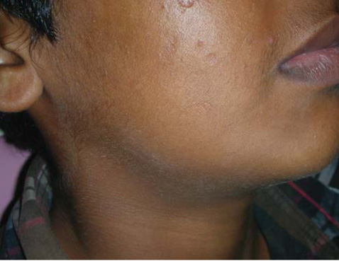
Fig. 1 Extraoral swelling is seen in the right submandibular region.
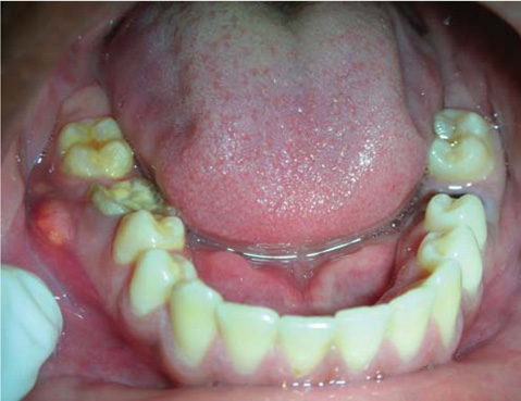
Fig. 2 Intraorally sinus opening is seen in relation to the right mandibular first molar.

Fig. 3 Mandibular occlusal radiograph shows bilateral cortical expansion along with trabeculation seen at the periphery of the lesion.
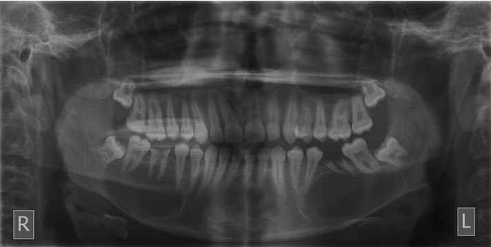
Fig. 4 Panoromic radiograph shows bilateral symmetrical unilocular radiolucencies with corticated borders.

Fig. 5 Histopathologic photograph shows arcading pattern of stratified squamous epithelium (H&E stain, ×40).
Reference
-
1. Regezi JA. Odontogenic cysts, odontogenic tumors, fibroosseous, and giant cell lesions of the jaws. Mod Pathol. 2002. 15:331–341.
Article2. Devenney-Cakir B, Subramaniam RM, Reddy SM, Imsande H, Gohel A, Sakai O. Cystic and cystic-appearing lesions of the mandible: review. AJR Am J Roentgenol. 2011. 196:WS66–WS77.3. Joshi NS, Sujan SG, Rachappa MM. An unusual case report of bilateral mandibular radicular cysts. Contemp Clin Dent. 2011. 2:59–62.
Article4. Shear M, Speight PM. Cysts of the oral and maxillofacial regions. 2007. 4th ed. Oxford: Blackwell;123–141.5. Bhaskar SN. Oral surgery - oral pathology conference No. 17, Walter Reed Army Medical Center. Periapical lesions - types, incidence, and clinical features. Oral Surg Oral Med Oral Pathol. 1966. 21:657–671.6. Syrjänen S, Tammisalo E, Lilja R, Syrjänen K. Radiological interpretation of the periapical cysts and granulomas. Dentomaxillofac Radiol. 1982. 11:89–92.
Article7. Regezi JA, Sciubba JJ, Jordan RC. Oral Pathology: clinical pathologic correlations. 2003. 4th ed. St Louis: WB Saunders;241–254.8. González-Alva P, Tanaka A, Oku Y, Yoshizawa D, Itoh S, Sakashita H, et al. Keratocystic odontogenic tumor: a retrospective study of 183 cases. J Oral Sci. 2008. 50:205–212.
Article9. Theodorou SJ, Theodorou DJ, Sartoris DJ. Imaging characteristics of neoplasms and other lesions of the jawbones: part 1. Odontogenic tumors and tumorlike lesions. Clin Imaging. 2007. 31:114–119.10. Dunfee BL, Sakai O, Pistey R, Gohel A. Radiologic and pathologic characteristics of benign and malignant lesions of the mandible. Radiographics. 2006. 26:1751–1768.
Article11. Hegde S, Shetty SR. Radiological features of familial Gorlin-Goltz syndrome. Imaging Sci Dent. 2012. 42:55–60.
Article12. Edwards PC, Fox J, Fantasia JE, Goldberg J, Kelsch RD. Bilateral central giant cell granulomas of the mandible in an 8-year-old girl with Noonan syndrome (Noonan-like/multiple giant cell lesion syndrome). Oral Surg Oral Med Oral Pathol Oral Radiol Endod. 2005. 99:334–340.
Article13. Ofluoglu D, Ertas B, Basarer N, Ergun S, Bilgic B, Tanyeri H. OC1 Bilateral central giant cell granuloma of the mandible: a case report. Oral Dis. 2006. 12:10.
Article14. Neyaz Z, Gadodia A, Gamanagatti S, Mukhopadhyay S. Radiographical approach to jaw lesions. Singapore Med J. 2008. 49:165–177.15. Jebasingh F, Jacob JJ, Shah A, Paul TV, Seshadri MS. Bilateral maxillary brown tumours as the first presentation of primary hyperparathyroidism. Oral Maxillofac Surg. 2008. 12:97–100.
Article16. Sham E, Leong J, Maher R, Schenberg M, Leung M, Mansour AK. Mandibular ameloblastoma: clinical experience and literature review. ANZ J Surg. 2009. 79:739–744.
Article17. Freitas DQ, Tempest LM, Sicoli E, Lopes-Neto FC. Bilateral dentigerous cysts: review of the literature and report of an unusual case. Dentomaxillofac Radiol. 2006. 35:464–468.
Article18. Gardner DG. Plexiform unicystic ameloblastoma: a diagnostic problem in dentigerous cysts. Cancer. 1981. 47:1358–1363.
Article19. MacDonald-Jankowski DS, Yeung R, Lee KM, Li TK. Ameloblastoma in the Hong Kong Chinese. Part 2: systematic review and radiological presentation. Dentomaxillofac Radiol. 2004. 33:141–151.
Article20. Lavery K, Blomquist JE, Awty MD, Stevens PJ. Squamous carcinoma arising in a dental cyst. Br Dent J. 1987. 162:259–260.
Article
- Full Text Links
-
- Actions
-
Cited
- CITED
-
- Close
- Share
-
- Similar articles
-
- Multiple jaw cysts not associated with basal cell nevus syndrome
- An Inflammatory Dentigerous Cyst Shows Rim Uptake on Bone Scan: A Case Report
- The Use of Endoscopy in Enucleation of Jaw Cysts
- Decompression Device Using a Stainless Steel Tube and Wire for Treatment of Odontogenic Cystic Lesions: A Technical Report
- Multiple Bilateral Thoracic Perineural Cysts: A Case Report
