Computed tomographic features of fibrous dysplasia of maxillofacial region
- Affiliations
-
- 1Department of Oral Medicine and Radiology, Nair Hospital Dental College, Mumbai, India. subodh204@yahoo.co.in
- 2Department of Oral Medicine and Radiology, Government Dental College and Hospital, Mumbai, India.
- KMID: 1974396
- DOI: http://doi.org/10.5624/isd.2011.41.1.23
Abstract
- PURPOSE
This study was to find the computed tomographic features of fibrous dysplasia of the maxillofacial region.
MATERIALS AND METHODS
All eight cases included in the study reported either to Government Dental College and Hospital or Nair Hospital Dental College, Mumbai between 2003 and 2009. The patients were prescribed computed tomogram in addition to conventional radiographs of maxillofacial region which were studied for characteristic features of fibrous dysplasia. The diagnosis of fibrous dysplasia was confirmed by histopathological report.
RESULTS
All cases showed the ill-defined margins of lesions except in the region where the lesions were extending to cortex of the involved bone. Internal structure of all cases showed ground glass appearance. Four cases of maxillary lesion showed the displacement of maxillary sinus maintaining the shape of maxillary sinus. Two cases showed complete obliteration of maxillary sinus. Displacement of inferior alveolar canal did not follow any typical pattern in any of the cases but was displaced in different directions.
CONCLUSION
The craniofacial type of fibrous dysplasia is as common as fibrous dysplasia of jaw. The margins, extent, internal structure and effect on surrounding structure are well detected on computed tomographic images.
MeSH Terms
Figure
-
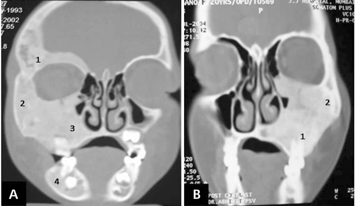
Fig. 1 CT images of fibrous dysplasia. A. Coronal section shows craniofacial type of fibrous dysplasia with involvement 1. frontal, temporal, 2. zygomatic, 3. maxilla, and 4. mandible. B. Another coronal section shows maxillofacial type with involvement of 1. maxilla and 2. zygomatic bone.
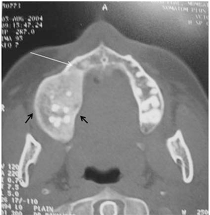
Fig. 2 Axial CT image shows ill-defined margin (white arrow) except in region where lesion is extending to the cortex (black double arrow).

Fig. 3 Coronal CT image shows lesion involving maxilla with increased density of maxilla giving ground glass appearance (black arrow).

Fig. 4 Coronal CT image shows maxillary sinus occupied with lesion. Note: the size is reduced but the shape of maxillary sinus is maintained (black arrow).

Fig. 5 CT images. A. Coronal section shows obliteration of 1. maxillary sinus. B. Oblique section shows complete obliteration of 1. maxillary and 2. sphenoid sinus.
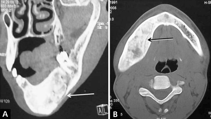
Fig. 6 CT images A. Oblique section shows displacement of inferior alveolar canal inferiorly (white arrow). B. Axial section showing displacement of inferior alveolar canal lingually (black arrow).
Reference
-
1. Bastepe M. The GNAS Locus: quintessential complex gene encoding Gsalpha, XLalphas, and other imprinted transcripts. Curr Genomics. 2007. 8:398–414.2. White SC, Pharoah MJ. Oral radiology: principles and interpretation. 2004. 5th ed. St. Louis: Mosby-Year Book Inc;485–491.3. Waldron CA. Neville BW, Damm DD, Allen CM, Bouquot JE, editors. Bone pathology. Oral and maxillofacial pathology. 1995. Philadelphia: WB Saunders;461–465.4. Daves ML, Yardley JH. Fibrous dysplasia of bone. Am J Med Sci. 1957. 234:590–606.
Article5. Eversole LR, Sabes WR, Rovin S. Fibrous dysplasia: a nosologic problem in the diagnosis of fibro-osseous lesions of the jaws. J Oral Pathol. 1972. 1:189–220.
Article6. MacDonald-Jankowski DS, Yeung R, Li TK, Lee KM. Computed tomography of fibrous dysplasia. Dentomaxillofac Radiol. 2004. 33:114–118.
Article7. Ricalde P, Horswell BB. Craniofacial fibrous dysplasia of the fronto-orbital region: a case series and literature review. J Oral Maxillofac Surg. 2001. 59:157–167.
Article8. Jacobsson S, Hallén O, Hollender L, Hansson CG, Lindström J. Fibro-osseous lesion of the mandible mimicking chronic osteomyelitis. Oral Surg Oral Med Oral Pathol. 1975. 40:433–444.
Article9. Posnick JC. Fibrous dysplasia of the craniomaxillofacial region: current clinical perspectives. Br J Oral Maxillofac Surg. 1998. 36:264–273.
Article10. MacDonald-Jankowski D. Fibrous dysplasia in the jaws of a Hong-Kong population: radiographic presentation and systematic review. Dentomaxillofac Radiol. 1999. 28:195–202.
Article11. Petrikowski CG, Pharoah MJ, Lee L, Grace MG. Radiographic differentiation of osteogenic sarcoma, osteomyelitis, and fibrous dysplasia of the jaws. Oral Surg Oral Med Oral Pathol Oral Radiol Endod. 1995. 80:744–750.
Article12. Akintoye SO, Lee JS, Feimster T, Booher S, Brahim J, Kingman A, et al. Dental characteristics of fibrous dysplasia and McCune-Albright syndrome. Oral Surg Oral Med Oral Pathol Oral Radiol Endod. 2003. 96:275–282.
Article
- Full Text Links
-
- Actions
-
Cited
- CITED
-
- Close
- Share
-
- Similar articles
-
- Fibrous dysplasia on left maxillofacial region
- Cervical Spinal Monostotic Fibrous Dysplasia: A Case Report
- Monostotic Fibrous Dysplasia of the Cervical Spine: A Case Report
- Radiographic Differential Diagnosis Between The Fibrous Dysplasia And The Ossifying Fibroma
- Pediculated Fibrous Dysplasia in Maxillary Sinus: A Case Report
