Metastatic Renal Cell Carcinoma in a Supraclavicular Lymph Node with No Known Primary: A Case Report
- Affiliations
-
- 1Department of Internal Medicine, Chungbuk National University College of Medicine, Cheongju, Korea. sook3529@hanamil.net
- 2Department of Pathology, Chungbuk National University College of Medicine, Cheongju, Korea.
Abstract
- Although metastasis is relatively frequent in cases of renal cell carcinoma (RCC), metastasis in the cervical or supraclavicular lymph node (LN) is relatively rare. Moreover, cases of metastatic RCC with a non-identifiable kidney mass are extremely rare. Here, the authors report a case of metastatic RCC in a supraclavicular LN without a primary kidney lesion. A 69-year-old man presented with a progressively enlarging right supraclavicular mass. Incisional biopsy of the affected supraclavicular LN was performed, and histological examination revealed metastatic RCC. However, no tumor was found in either kidney, despite various examinations. The patient was treated with radiotherapy followed by sunitinib. After three months on sunitinib, a follow-up computed tomography scan revealed that the supraclavicular LN had markedly decreased, and after 20 months, the disease had not progressed. This case suggests that, even when there is no primary kidney lesion, clinicians must consider the possibility of metastatic RCC when evaluating patients with clear cell carcinoma with an unknown primary site.
Keyword
MeSH Terms
Figure
-
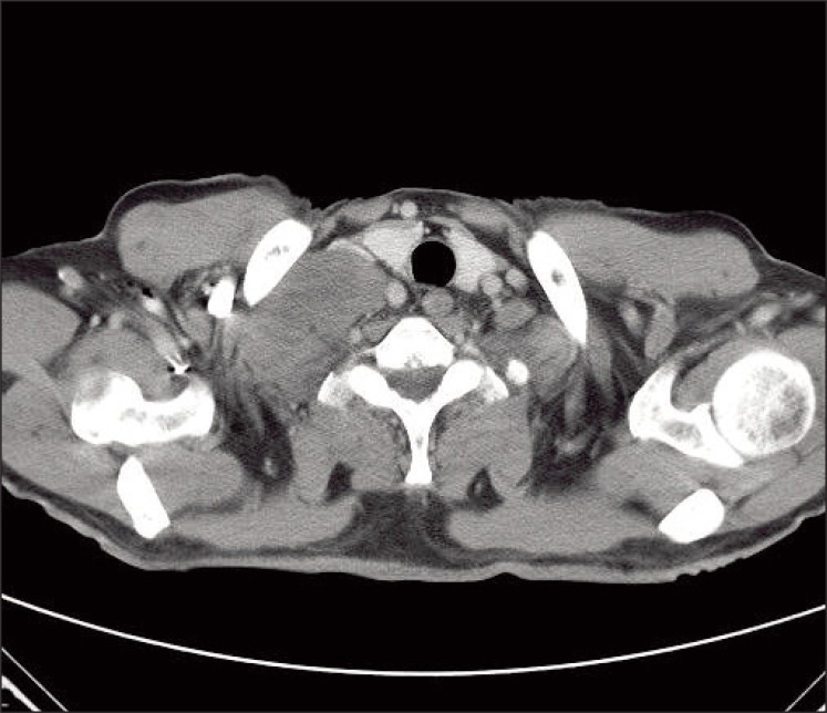
Fig. 1 Computed tomography of the neck showing lymph node enlargement in the right supraclavicular area.
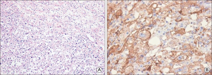
Fig. 2 Pathological features of an incisional biopsy of the supraclavicular lymph node. (A) The tumor cells had clear cytoplasm and vesicular nuclei with prominent nucleoli, and were are arranged in nests admixed with intervening blood vessels (H&E staining, ×200). (B) Immunohistochemistry showed that the tumor cells were positive for CD10 (×400).

Fig. 3 Computed tomography (CT) of the abdomen/pelvis and positron emission tomography (PET)-CT. (A) CT of the abdomen and pelvis revealing no tumor in either kidney. (B) PET-CT images show the right supraclavicular lymph node with 18-fluoro-deoxyglucose (FDG) uptake (standardized uptake value, SUV, 14.7), compatible with malignant lymphadenopathy. However, no other FDG uptake was found in either kidney, and there were no additional metastatic lesions.
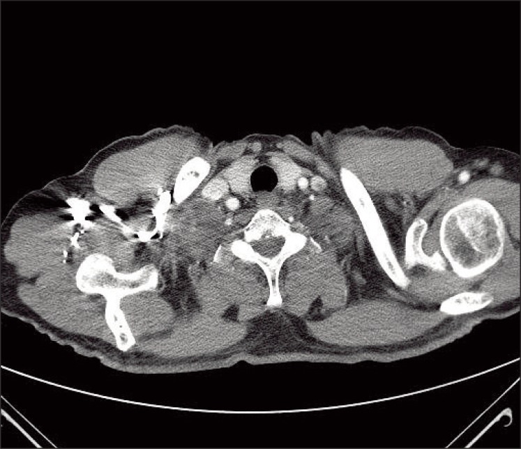
Fig. 4 Computed tomography of the neck after radiotherapy and two cycles of sunitinib showing markedly reduced lymph node enlargement in the right supraclavicular area.
Reference
-
1. Golimbu M, Joshi P, Sperber A, Tessler A, Al-Askari S, Morales P. Renal cell carcinoma: survival and prognostic factors. Urology. 1986; 27:291–301. PMID: 3962052.
Article2. Maldazys JD, deKernion JB. Prognostic factors in metastatic renal carcinoma. J Urol. 1986; 136:376–379. PMID: 3735498.
Article3. Pritchyk KM, Schiff BA, Newkirk KA, Krowiak E, Deeb ZE. Metastatic renal cell carcinoma to the head and neck. Laryngoscope. 2002; 112:1598–1602. PMID: 12352670.
Article4. Cheng ET, Greene D, Koch RJ. Metastatic renal cell carcinoma to the nose. Otolaryngol Head Neck Surg. 2000; 122:464. PMID: 10699832.
Article5. Miyamoto R, Helmus C. Hypernephroma metastatic to the head and neck. Laryngoscope. 1973; 83:898–905. PMID: 4711327.
Article6. Kawakita M, Kawamura J, Hida S, Ooishi K, Okada K, Yoshida O. Renal cell carcinoma, primary lesion which was not easily identified: report of two cases. Hinyokika Kiyo. 1985; 31:463–473. PMID: 4025083.7. Bhatia S, Ng S, Hodder SC. Metastatic cutaneous head and neck renal cell carcinoma with no known primary: case report. Br J Oral Maxillofac Surg. 2010; 48:214–215. PMID: 20036041.
Article8. Rasco DW, Assikis V, Marshall F. Integrating metastasectomy in the management of advanced urological malignancies-where are we in 2005? J Urol. 2006; 176:1921–1926. PMID: 17070212.
Article9. Mendel DB, Laird AD, Xin X, Louie SG, Christensen JG, Li G, et al. In vivo antitumor activity of SU11248, a novel tyrosine kinase inhibitor targeting vascular endothelial growth factor and platelet-derived growth factor receptors: determination of a pharmacokinetic/pharmacodynamic relationship. Clin Cancer Res. 2003; 9:327–337. PMID: 12538485.
- Full Text Links
-
- Actions
-
Cited
- CITED
-
- Close
- Share
-
- Similar articles
-
- Metastatic tumors in supraclavicular lymph node: pathological analysis of 125 cases
- A case of metastatic squamous cell carcinoma of the mediastinum with unknown primary tumor
- Indolent Metastatic Squamous Cell Carcinoma of Unknown Primary in the Intrathoracic Lymph Node: A Case Report and Review of the Literatures
- A case of minimal uterine serous carcinoma with distant lymph node metastasis without peritoneal dissemination
- Mesothelial Cell Inclusions Mimicking Metastatic Carcinoma in Mediastinal Lymph Node: A Case Report
