Tuberc Respir Dis.
2006 Dec;61(6):547-553. 10.4046/trd.2006.61.6.547.
Comparison of Thinprep (Liquid-Based Cytology) and Conventional Cytology: Abnormal Lesion on Bronchoscopy
- Affiliations
-
- 1Department of Internal Medicine, College of Medicine, Konyang University, Daejeon, Korea. sk1609@hanmail.net
- 2Department of Pathology, College of Medicine, Konyang University, Daejeon, Korea.
- KMID: 1970239
- DOI: http://doi.org/10.4046/trd.2006.61.6.547
Abstract
-
BACKGROUND: Liquid-based cytology is currently known as an effective method, and cervical cytology has been shown to be especially effective from of malignancy detection. In our study, the cytological detection rates of the Thinprep (Liquid-based cytology) and conventional cytology (bronchial washing & brushing) for endobronchial lesions were compared.
METHODS
Between July 2005 and September 2005, the data from 30 patients with respiration symptom, who had shown abnormal lesion on bronchoscopy, were collected.
RESULTS
The bronchoscopic biopsy group was consisted of 30 cytodiagnosis specimens, 24 of which were confirmed to be malignant. The others were tuberculosis (4), bronchiectasis and bronchopulmonary fistula (1 each). Of the 24 malignant case, cancer or atypical cells were detected in 19, 17 and 12 of the Thinprep, brushing cytology and washing cytology cases, respectively. None one of the methods detected cancer cells in the non-malignant specimens. Washing cytology has shown sensitivity, specificity, and positive and negative predictive values of 50, 100, 100 and 33.3% respectively. Brushing cytology has shown sensitivity, specificity, and positive and negative predictive values of 70.8, 100, 100 and 46.2%, respectively. Thinprep has shown sensitivity, specificity, and positive and negative predictive values of 79.2, 100, 100 and 54%, respectively.
CONCLUSIONS
Thinprep (liquid-based cytology) showed better sensitivity and negative predictive values for the evaluation of lung cancer than conventional cytology. However a large-scale study will be needed in the future.
MeSH Terms
Figure
-
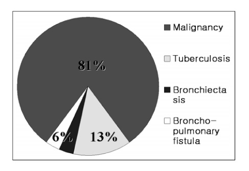
Figure 1 Final diagnosis of the patient.

Figure 2 Cellularity.

Figure 3 Preservation.

Figure 4 Background.

Figure 5 Sensitivity.

Figure 6 Negative predictability.

Figure 7 Squamous cell carcinoma (a) Conventional preparation (b) Thinprep.(Papanicolaou stain, ×200)
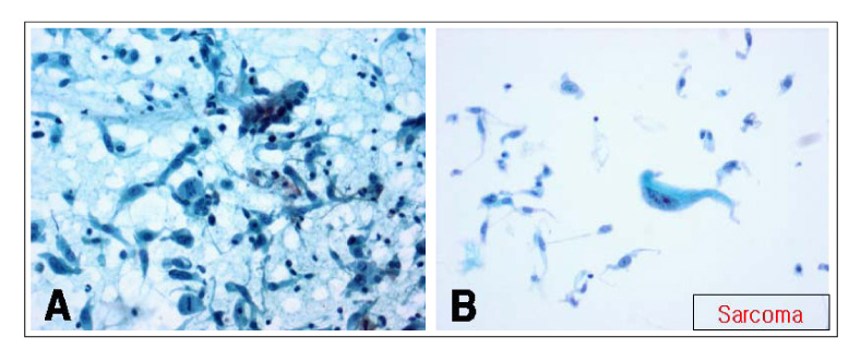
Figure 8 Sarcoma (a) Conventional preparation (b) ThinPrep (Papanicolaou stain, ×200)
Cited by 1 articles
-
Comparison of ThinPrep Cytology and Conventional Cytology in Bronchial Washing for Lung Cancer
Jungsuk An, Hyun Yee Cho, Dong Hae Chung, Seung Yeon Ha
J Lung Cancer. 2012;11(2):84-88. doi: 10.6058/jlc.2012.11.2.84.
Reference
-
1. Arroliga AC, Matthay RA. The role of bronchoscopy in lung cancer. Clin Chest Med. 1993. 14:87–98.2. Linder J. Recent advances in thin-layer cytology. Diagn Cytopathol. 1998. 18:24–32.3. Compton J, Boyle E, Barck M, Jicha D, Reale FR. Sputum cytology: a comparison of two methods, the conventional saccamanno preparation and the ThinPrep Processor [Abstract]. Acta Cytol. 1993. 37:797.4. Miller FS, Pizzo CJ. Cytospin vs. thin-layer technology: nongynecologic evaluation [Abstract]. Acta Cytol. 1993. 37:798.5. Burroughs FN, Erozan YS. Comparison of ThinPrep and conventional preparation in alimentary tract cytopathology [Abstract]. Acta Cytol. 1993. 37:801.6. Kish JK, Vallera DU, Ruby SG. Comparative study of nongynecologic processing by ThinPrep vs. conventional methodology: ratinale for the use of ThinPrep [Abstract]. Acta Cytol. 1993. 37:801.7. Papillo JL, Lee KR, Manna EA. Clinical evlauation of the ThinPrep method for the preparation of nongynecologic material. Acta Cytol. 1992. 36:651–652.8. Dudgeon LS, Wrigley CH. On the demonstration of particles of malignant growth in the sputum by means of wet-film metho. J Laryngol Otol. 1935. 50:752.9. Herbut PA, Clerf LH. Bronchogenic carcinoma: diagnosis by cytologic study of bronchoscopically removed secretions. JAMA. 1946. 130:1006.10. Kvale PA, Bode FR, Kini S. Diagnostic accuracy in lung cancer: comparison of techniques used in association with flexible fiberoptic bronchoscopy. Chest. 1976. 69:752–757.11. Funahashi A, Browne TK, Houser WC, Hranicka LJ. Diagnostic value of bronchial aspirate and postbronchoscopic sputum in fiberoptic bronchoscopy. Chest. 1979. 76:514–517.12. Mak VH, Johnston ID, Hetzel MR, Grubb C. Value of washings and brushings at fiberoptic bronchoscopy in the diagnosis of lung cancer. Thorax. 1990. 45:373–376.13. Struve-Christensen E, Michaelsen M, Mossing N. The diagnostic value of bronchial washing in lung cancer. J Thorac Cardiovasc Surg. 1974. 68:313–317.14. Kurtycz DF, Hoerl HD. Thin-layer technology: tempered enthusiasm. Diagn Cytopathol. 2000. 23:1–5.15. Cytyc Corporation. Operator's manaual: ThinPrep Processor. 1993. Marlborough, MA: Cytyc Corperation.16. Fischler DF, Toddy SM. Nongynecologic cytology utilizing the ThinPrep Precessor. Acta Cytol Au. 1996. 40:669–675.17. Leung CS, Chiu B, Bell V. Comparison of ThinPrep and conventional preparations. Diagn Cytopathol. 1997. 16:368–371.18. Papillo JL, Lapen D. Cell yield: ThinPrep vs. cytocentrifuge. Acta Cytol. 1994. 38:33–36.19. Hees K, Lebeau PB. Cpmparison of conventional and ThinPrep preparations of mucoid cytology samples. Diagn Cytopathol. 1995. 12:181–185.20. Rana DN, O'Donnell M, Malkin A, Griffin M. A comparative study: conventional preparation and ThinPrep 2000 in respiratory cytology. Cytopathology. 2001. 12:390–398.21. Hoerl HD, Schink J, Hartenbach E, Wagner JL, Kurtycz DF. Exfoliative cytology of promary poorly diifferentiated (small-cell) neuroendocrine carcinoma of the uterine cervix in ThinPrep material: a case report. Diagn Cytopathol. 2000. 23:14–18.22. Michael CW, Hunter B. Interpretation of fine-needle aspirates precessed by the ThinPrep technique: cytologic artifacts diagnostic pitfalls. Diagn Cytopathol. 2000. 23:6–13.
- Full Text Links
-
- Actions
-
Cited
- CITED
-
- Close
- Share
-
- Similar articles
-
- Comparison of ThinPrep Cytology and Conventional Cytology in Bronchial Washing for Lung Cancer
- Diagnostic Value of Urine Cytology in 236 cases; a Comparison of Liquid-Based Preparation and Conventional Cytospin Method
- A Comparison of Conventional Cytology and ThinPrep Cytology of Bronchial Washing Fluid in the Diagnosis of Lung Cancer
- A Comparison Between ThinPrep Monolayer and Cytospin Cytology for the Detection of Bladder Cancer
- Detecting Malignant Urothelial Cells by Morphometric Analysis of ThinPrep(R) Liquid-based Urine Cytology Specimens
