Bilateral Metachronous Breast Cancer with Bilateral Recurrences: A Case Report and Literature Review
- Affiliations
-
- 1Department of Radiology, Kyung Hee University Hospital, College of Medicine, Kyung Hee University, Seoul, Korea. sonyumee@naver.com
- 2Department of Radiology, Research Institute of Radiological Science, Yonsei University College of Medicine, Seoul, Korea.
- KMID: 1941784
- DOI: http://doi.org/10.3348/jksr.2014.70.5.369
Abstract
- The incidence of bilateral breast cancer has been reported to range from 0.4% to 14%, and it increases gradually as a result of improved early detection capabilities and longer survival times. We report a rare case where the bilateral breast cancers occurred as a metachronous bilateral breast cancer with bilateral recurrences, detected by mammography, and the rapid growth of tumor that manifested as microcalcification and skin thickening within 3 months.
Figure
-

Fig. 1 Mammography of 51-year-old woman with a history of bilateral partial mastectomy. A. A screening mammogram of the right breast showed a few punctuate calcifications in a diffuse distribution categorized as category 2 (benign finding). The patient revisited the hospital after 3 months and complained of right breast swelling. B. A follow-up mammogram after 3 months showed developing pleomorphic microcalcifications (solid arrow) in a segmental distribution and skin thickening (arrowheads). C. Spot magnification view showed apparently pleomorphic microcalcifications with a segmental distribution (solid arrow) extending to the right subareolar area (dotted arrow), prompting a biopsy and the result of biopsy was invasive ductal carcinoma.
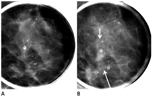
Fig. 2 Follow-up left spot magnification view during surveillance. A. Spot magnification of the left breast showed a few punctuate microcalcifications and macrocalcifications, which were categorized as category 3 (probably benign finding). B. Follow-up spot magnification view after 6 months showed an increasing number of suspicious pleomorphic microcalcifications (solid arrows).
Reference
-
1. Takahashi H, Watanabe K, Takahashi M, Taguchi K, Sasaki F, Todo S. The impact of bilateral breast cancer on the prognosis of breast cancer: a comparative study with unilateral breast cancer. Breast Cancer. 2005; 12:196–202.2. Donovan AJ. Bilateral breast cancer. Surg Clin North Am. 1990; 70:1141–1149.3. Dawson LA, Chow E, Goss PE. Evolving perspectives in contralateral breast cancer. Eur J Cancer. 1998; 34:2000–2009.4. American College of Radiology. ACR BI-RADS: Mammography. ACR Breast Imaging Reporting and Data System, Breast Imaging Atlas. 4th ed. Reston, VA: American College of Radiology;2003.5. Heron DE, Komarnicky LT, Hyslop T, Schwartz GF, Mansfield CM. Bilateral breast carcinoma: risk factors and outcomes for patients with synchronous and metachronous disease. Cancer. 2000; 88:2739–2750.6. Lou L, Cong XL, Yu GF, Li JC, Ma YX. US findings of bilateral primary breast cancer: retrospective study. Eur J Radiol. 2007; 61:154–157.7. Lee JS, Grant CS, Donohue JH, Crotty TB, Harmsen WS, Ilstrup DM. Arguments against routine contralateral mastectomy or undirected biopsy for invasive lobular breast cancer. Surgery. 1995; 118:640–647.8. Ng AK, Kenney LB, Gilbert ES, Travis LB. Secondary malignancies across the age spectrum. Semin Radiat Oncol. 2010; 20:67–78.9. Cowan WK, Angus B, Gray JC, Lunt LG, al-Tamimi SR. A study of interval breast cancer within the NHS breast screening programme. J Clin Pathol. 2000; 53:140–146.10. Robertson C, Ragupathy SK, Boachie C, Fraser C, Heys SD, Maclennan G, et al. Surveillance mammography for detecting ipsilateral breast tumour recurrence and metachronous contralateral breast cancer: a systematic review. Eur Radiol. 2011; 21:2484–2491.
