Coronary Artery Anomalies: Assessment with Electrocardiography-Gated Multidetector-Row CT at a Single Center in Korea
- Affiliations
-
- 1Department of Radiology, Ajou University School of Medicine, Suwon, Korea. kdklsm@ajou.ac.kr
- 2Department of Cardiology, Ajou University School of Medicine, Suwon, Korea.
- KMID: 1941753
- DOI: http://doi.org/10.3348/jksr.2015.72.4.207
Abstract
- PURPOSE
To determine the prevalence of coronary anomalies using coronary computed tomography angiography (CCTA) and to evaluate the relationship between coronary artery anomalies and chest pain.
MATERIALS AND METHODS
A total of 12676 patients underwent CCTA scans at our institution between December 2006 and April 2013 using a 64-slice CT and a 128-slice dual-source CT. We determined the prevalence of coronary artery anomalies according to the classification system proposed by Greenberg. The presence or absence of chest pain with each coronary artery anomaly was also evaluated.
RESULTS
Coronary anomalies were found in 176 patients (1.39%) at our institute. Anomalies of origination, course, and termination were detected in 118 (0.93%), 28 (0.22%), and 30 (0.24%) patients, respectively. After the exclusion of 32 patients with combined heart disease, typical (n = 16; 11.1%) or atypical (n = 28; 19.4%) chest pain was present in 44 (30.6%) of the 144 patients at the time of diagnosis.
CONCLUSION
The prevalence of coronary artery anomalies was 1.39% at our hospital. After the exclusion of patients with combined heart disease, 11.1% had typical chest pain at the time of diagnosis.
MeSH Terms
Figure
-

Fig. 1 63 years old male patient with atypical angina. A, B. Volume rendering technique and three-dimensional maximum intensity projection images show aplasia of right coronary artery (RCA) and collateral vessel from left circumflex artery (arrow). C. Coronary angiography shows left dominant coronary artery with RCA aplasia.
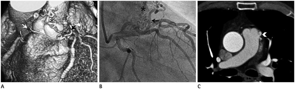
Fig. 2 82 years old male patient with atypical angina. A, B. Volume rendering technique and coronary angiography show tortuous vessels arising from the proximal left anterior descending (black arrow) and right coronary sinus (white arrow) and draining into pulmonary trunk. Multiple tortuous vascular structures passes anterior to the pulmonary artery and forms a network (*) before it enters the pulmonary trunk. C. Axial CT image shows the entry site (arrowhead) into pulmonary trunk.

Fig. 3 57 years old female patient with typical angina. A. Volume rendering technique shows anomalous right coronary artery (RCA) ostium from the left coronary sinus. B. Oblique sagittal multiplanar reformation image shows RCA (arrow) travels between aorta (Ao) and pulmonary artery (*).
Reference
-
1. Alexander RW, Griffith GC. Anomalies of the coronary arteries and their clinical significance. Circulation. 1956; 14:800–805.2. Angelini P, Velasco JA, Flamm S. Coronary anomalies: incidence, pathophysiology, and clinical relevance. Circulation. 2002; 105:2449–2454.3. Schmitt R, Froehner S, Brunn J, Wagner M, Brunner H, Cherevatyy O, et al. Congenital anomalies of the coronary arteries: imaging with contrast-enhanced, multidetector computed tomography. Eur Radiol. 2005; 15:1110–1121.4. Duran C, Kantarci M, Durur Subasi I, Gulbaran M, Sevimli S, Bayram E, et al. Remarkable anatomic anomalies of coronary arteries and their clinical importance: a multidetector computed tomography angiographic study. J Comput Assist Tomogr. 2006; 30:939–948.5. Srinivasan KG, Gaikwad A, Kannan BR, Ritesh K, Ushanandini KP. Congenital coronary artery anomalies: diagnosis with 64 slice multidetector row computed tomography coronary angiography: a single-centre study. J Med Imaging Radiat Oncol. 2008; 52:148–154.6. Tariq R, Kureshi SB, Siddiqui UT, Ahmed R. Congenital anomalies of coronary arteries: diagnosis with 64 slice multidetector CT. Eur J Radiol. 2012; 81:1790–1797.7. Greenberg MA, Fish BG, Spindola-Franco H. Congenital anomalies of the coronary arteries. Classification and significance. Radiol Clin North Am. 1989; 27:1127–1146.8. Dodge-Khatami A, Mavroudis C, Backer CL. Congenital Heart Surgery Nomenclature and Database Project: anomalies of the coronary arteries. Ann Thorac Surg. 2000; 69:4 Suppl. S270–S297.9. Kim SY, Seo JB, Do KH, Heo JN, Lee JS, Song JW, et al. Coronary artery anomalies: classification and ECG-gated multi-detector row CT findings with angiographic correlation. Radiographics. 2006; 26:317–333. discussion 333-334.10. Earls JP. Coronary artery anomalies. Tech Vasc Interv Radiol. 2006; 9:210–217.11. Kang JW, Seo JB, Chae EJ, Jang YM, Do KH, Lee JS, et al. Coronary artery anomalies: classification and electrocardiogram-gated multidetector computed tomographic findings. Semin Ultrasound CT MR. 2008; 29:182–194.12. Patel S. Normal and anomalous anatomy of the coronary arteries. Semin Roentgenol. 2008; 43:100–112.13. Sundaram B, Kreml R, Patel S. Imaging of coronary artery anomalies. Radiol Clin North Am. 2010; 48:711–727.14. Pursnani A, Jacobs JE, Saremi F, Levisman J, Makaryus AN, Capuñay C, et al. Coronary CTA assessment of coronary anomalies. J Cardiovasc Comput Tomogr. 2012; 6:48–59.15. van Ooijen PM, Dorgelo J, Zijlstra F, Oudkerk M. Detection, visualization and evaluation of anomalous coronary anatomy on 16-slice multidetector-row CT. Eur Radiol. 2004; 14:2163–2171.16. Shi H, Aschoff AJ, Brambs HJ, Hoffmann MH. Multislice CT imaging of anomalous coronary arteries. Eur Radiol. 2004; 14:2172–2181.17. Karaca M, Kirilmaz A, Oncel G, Oncel D, Yilmaz H, Tamci B, et al. Contrast-enhanced 64-slice computed tomography in detection and evaluation of anomalous coronary arteries. Tohoku J Exp Med. 2007; 213:249–259.18. Shriki JE, Shinbane JS, Rashid MA, Hindoyan A, Withey JG, DeFrance A, et al. Identifying, characterizing, and classifying congenital anomalies of the coronary arteries. Radiographics. 2012; 32:453–468.19. Tresoldi S, Mezzanzanica M, Campari A, Salerno Uriarte D, Cornalba G. Role of computed tomography coronary angiography in the management of coronary anomalies. J Card Surg. 2013; 28:33–36.20. Gibbons RJ, Balady GJ, Beasley JW, Bricker JT, Duvernoy WF, Froelicher VF, et al. ACC/AHA guidelines for exercise testing: executive summary. A report of the American College of Cardiology/American Heart Association Task Force on Practice Guidelines (Committee on Exercise Testing). Circulation. 1997; 96:345–354.21. Diamond GA. A clinically relevant classification of chest discomfort. J Am Coll Cardiol. 1983; 1(2 Pt 1):574–575.22. Möhlenkamp S, Hort W, Ge J, Erbel R. Update on myocardial bridging. Circulation. 2002; 106:2616–2622.23. Zeina AR, Odeh M, Blinder J, Rosenschein U, Barmeir E. Myocardial bridge: evaluation on MDCT. AJR Am J Roentgenol. 2007; 188:1069–1073.24. Konen E, Goitein O, Sternik L, Eshet Y, Shemesh J, Di Segni E. The prevalence and anatomical patterns of intramuscular coronary arteries: a coronary computed tomography angiographic study. J Am Coll Cardiol. 2007; 49:587–593.25. Ko SM, Choi JS, Nam CW, Hur SH. Incidence and clinical significance of myocardial bridging with ECG-gated 16-row MDCT coronary angiography. Int J Cardiovasc Imaging. 2008; 24:445–452.26. Kim SY, Lee YS, Lee JB, Ryu JK, Choi JY, Chang SG, et al. Evaluation of myocardial bridge with multidetector computed tomography. Circ J. 2010; 74:137–141.27. Montaudon M, Latrabe V, Iriart X, Caix P, Laurent F. Congenital coronary arteries anomalies: review of the literature and multidetector computed tomography (MDCT)-appearance. Surg Radiol Anat. 2007; 29:343–355.28. Erol C, Seker M. The prevalence of coronary artery variations on coronary computed tomography angiography. Acta Radiol. 2012; 53:278–284.29. Lee HJ, Hong YJ, Kim HY, Lee J, Hur J, Choi BW, et al. Anomalous origin of the right coronary artery from the left coronary sinus with an interarterial course: subtypes and clinical importance. Radiology. 2012; 262:101–108.30. Desmet W, Vanhaecke J, Vrolix M, Van de Werf F, Piessens J, Willems J, et al. Isolated single coronary artery: a review of 50,000 consecutive coronary angiographies. Eur Heart J. 1992; 13:1637–1640.31. Yamanaka O, Hobbs RE. Coronary artery anomalies in 126,595 patients undergoing coronary arteriography. Cathet Cardiovasc Diagn. 1990; 21:28–40.32. Kimbiris D, Iskandrian AS, Segal BL, Bemis CE. Anomalous aortic origin of coronary arteries. Circulation. 1978; 58:606–615.33. Donaldson RM, Raphael M, Radley-Smith R, Yacoub MH, Ross DN. Angiographic identification of primary coronary anomalies causing impaired myocardial perfusion. Cathet Cardiovasc Diagn. 1983; 9:237–249.34. Chaitman BR, Lespérance J, Saltiel J, Bourassa MG. Clinical, angiographic, and hemodynamic findings in patients with anomalous origin of the coronary arteries. Circulation. 1976; 53:122–131.35. Bunce NH, Lorenz CH, Keegan J, Lesser J, Reyes EM, Firmin DN, et al. Coronary artery anomalies: assessment with free-breathing three-dimensional coronary MR angiography. Radiology. 2003; 227:201–208.36. Ropers D, Gehling G, Pohle K, Maeffert R, Regenfus M, Moshage W, et al. Images in cardiovascular medicine. Anomalous course of the left main or left anterior descending coronary artery originating from the right sinus of valsalva: identification of four common variations by electron beam tomography. Circulation. 2002; 105:e42–e43.37. Cheitlin MD, De Castro CM, McAllister HA. Sudden death as a complication of anomalous left coronary origin from the anterior sinus of Valsalva, A not-so-minor congenital anomaly. Circulation. 1974; 50:780–787.38. Roberts WC, Siegel RJ, Zipes DP. Origin of the right coronary artery from the left sinus of valsalva and its functional consequences: analysis of 10 necropsy patients. Am J Cardiol. 1982; 49:863–868.39. Virmani R, Chun PK, Goldstein RE, Robinowitz M, McAllister HA. Acute takeoffs of the coronary arteries along the aortic wall and congenital coronary ostial valve-like ridges: association with sudden death. J Am Coll Cardiol. 1984; 3:766–771.40. Kragel AH, Roberts WC. Anomalous origin of either the right or left main coronary artery from the aorta with subsequent coursing between aorta and pulmonary trunk: analysis of 32 necropsy cases. Am J Cardiol. 1988; 62(10 Pt 1):771–777.41. Frescura C, Basso C, Thiene G, Corrado D, Pennelli T, Angelini A, et al. Anomalous origin of coronary arteries and risk of sudden death: a study based on an autopsy population of congenital heart disease. Hum Pathol. 1998; 29:689–695.42. Lee BY. Anomalous right coronary artery from the left coronary sinus with an interarterial course: is it really dangerous? Korean Circ J. 2009; 39:175–179.43. Namgung J, Kim JA. The prevalence of coronary anomalies in a single center of Korea: origination, course, and termination anomalies of aberrant coronary arteries detected by ECG-gated cardiac MDCT. BMC Cardiovasc Disord. 2014; 14:48.44. Spindola-Franco H, Grose R, Solomon N. Dual left anterior descending coronary artery: angiographic description of important variants and surgical implications. Am Heart J. 1983; 105:445–455.45. Gowda RM, Vasavada BC, Khan IA. Coronary artery fistulas: clinical and therapeutic considerations. Int J Cardiol. 2006; 107:7–10.46. Zenooz NA, Habibi R, Mammen L, Finn JP, Gilkeson RC. Coronary artery fistulas: CT findings. Radiographics. 2009; 29:781–789.47. Braden DS, O'Neal KR, McMullan MR, Ebeid MR. Congenital coronary arteriovenous fistula presenting with syncope. Pediatr Cardiol. 2002; 23:218–220.48. Lee CM, Leung TK, Wang HJ, Lee WH, Shen LK, Chen YY. Identification of a coronary-to-bronchial-artery communication with MDCT shows the diagnostic potential of this new technology: case report and review. J Thorac Imaging. 2007; 22:274–276.
- Full Text Links
-
- Actions
-
Cited
- CITED
-
- Close
- Share
-
- Similar articles
-
- A case of coronary artery-pulmonary artery fistula communicated with aorto-pulmonary fistula via common channel detected by Multidetector row CT (MDCT) and coronary angiography
- A Single Coronary Artery: Right Coronary Artery Originating From the Distal Left Circumflex Artery
- Technical Aspect of Coronary CT Angiography: Imaging Tips and Safety Issues
- Unusual Coronary Artery Fistula: Left Anterior Descending Coronary Artery - Left Ventricular Fistula Diagnosed by ECG-Gated Multi-Detector Row Coronary CT Angiography
- Coronary Artery Anomaly, What Radiologist Should Know?
