Giant Cell Reparative Granuloma in Soft Tissue of Foot: A Case Report
- Affiliations
-
- 1Department of Radiology, Sanggye Paik Hospital, Inje University College of Medicine, Seoul, Korea. merita@paik.ac.kr
- 2Department of Pathology, Sanggye Paik Hospital, Inje University College of Medicine, Seoul, Korea.
- KMID: 1897263
- DOI: http://doi.org/10.3348/jksr.2014.70.1.65
Abstract
- Giant cell reparative granuloma is a benign reactive process following intraosseous hemorrhage rather than a true tumor. This lesion most commonly affects the maxilla and mandible, followed by phalanges, hands, and feet. Local invasion of surrounding soft tissue is a typical feature of giant cell reparative granuloma in the bones of the upper and lower limbs. We present the rare case of giant cell reparative granuloma arising from soft tissue of the foot without erosion or engulfing of the adjacent bone.
Figure
-
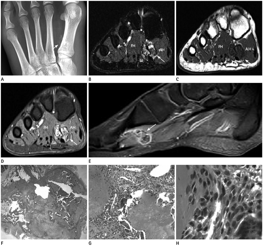
Fig. 1 Giant cell reparative granuloma of soft tissue of foot in 18-year-old woman. A. Plain radiograph shows clustered and punctate soft tissue calcifications around base of first metatarsal bone (arrow). B, C. Coronal MR images reveal a moderately defined intermuscular mass (arrows) between flexor hallucis brevis (FH), lumbricallis (Lu), and adductor hallucis (AH) muscle, showing heterogeneous high signal intensity on fat-suppressed T2-weighted image (B) and mixed intermediate and low signal intensity on T1-weighted image (C). D, E. Coronal (D) and sagittal (E) gadolineum enhanced fat-suppressed T1-weighted images reveal heterogeneous enhancement of the lesion (arrows). F-H. Photomicrograph of surgical specimen shows embedded bony mass in skeletal muscle and fatty tissue (H&E stain, × 10) (F), consists of fibrous stromal tissue in reactive bone formation (abundant osteochondral matrix) (H&E stain, × 100) (G) and spindle cell (fibroblasts) without atypia, arranged in a whorled to fascicular pattern and occasionally admixed osteoclastic giant cells (H&E stain, × 400) (H).
Reference
-
1. Jaffe HL. Giant-cell reparative granuloma, traumatic bone cyst, and fibrous (fibro-oseous) dysplasia of the jawbones. Oral Surg Oral Med Oral Pathol. 1953; 6:159–175.2. Felsberg GJ, Tien RD, McLendon RE. Frontoethmoidal giant cell reparative granuloma. AJNR Am J Neuroradiol. 1995; 16:1551–1554.3. Morris JM, Lane JI, Witte RJ, Thompson DM. Giant cell reparative granuloma of the nasal cavity. AJNR Am J Neuroradiol. 2004; 25:1263–1265.4. Murphey MD, Nomikos GC, Flemming DJ, Gannon FH, Temple HT, Kransdorf MJ. From the archives of AFIP. Imaging of giant cell tumor and giant cell reparative granuloma of bone: radiologic-pathologic correlation. Radiographics. 2001; 21:1283–1309.5. Eck HC, Weiner SD. Giant-cell reparative granuloma of the hands and feet. Orthopedics. 2004; 27:67–70.6. Nackos JS, Wiggins RH 3rd, Harnsberger HR. CT and MR imaging of giant cell granuloma of the craniofacial bones. AJNR Am J Neuroradiol. 2006; 27:1651–1653.7. Parikh J, Hyare H, Saifuddin A. The imaging features of post-traumatic myositis ossificans, with emphasis on MRI. Clin Radiol. 2002; 57:1058–1066.8. Gartner L, Pearce CJ, Saifuddin A. The role of the plain radiograph in the characterisation of soft tissue tumours. Skeletal Radiol. 2009; 38:549–558.
- Full Text Links
-
- Actions
-
Cited
- CITED
-
- Close
- Share
-
- Similar articles
-
- MR Imaging of Uncommon Soft Tissue Tumors in the Foot: A Pictorial Essay
- Giant Cell Reparative Granuloma in the Temporal Bone of a 4-Month-Old Infant
- A Case of Papular Elastolytic Giant Cell Granuloma on the Knees
- Ossified Soft Tissue Recurrence of Giant Cell Tumor: Three Case Report
- A Case of the Giant Cell Reaction in the Foot
